Return to Case List with Diagnoses or Case List as Unknowns

SPECT ARTIFACT
Authored By: Joanna Fair and Barry Siegel, Prof of Radiology.
Patient: 64 year old
History: Elevated 6-HIAA.
Image Size:[small][as-submitted]

Multimedia: 129392_1_submitted.avi
Maximum-intensity-projection (MIP) images of the processed SPECT data from an In-111 pentetreotide scan, obtained 24 hours post injection.

Fig. 2
Same reconstructed SPECT data, displayed as transaxial images.

Multimedia: 129392_3_submitted.avi
Raw data (projection images) from the same scan.

Multimedia: 129392_4_submitted.avi
Subset (few frames) of projection data showing the data collapse into a single point before disappearing.

Multimedia: 129392_5_submitted.avi
Repeat imaging of the same patient acquired 2 hours later. Maximum- intensity-projection (MIP) images are shown.
Image Size:[small][as-submitted]

Findings:

Radiopharmaceutical: 6 mCi In-111 pentetreotide i.v.


Please review Fig. 1 and Fig. 2.


[pagebreak]

There is a rod-like area of apparent increased activity on the SPECT maximum-intensity projection (MIP) images (Fig. 1).  The transaxial filtered-back-projection SPECT images show a corresponding linear streak of increased activity (Fig. 2).

What would you do next?
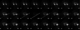
[pagebreak]

Examination of the raw projection images (Fig. 3) shows absent data for the last approximately 20 frames of the acquisition.  Focusing on just the last few frames with data (Fig. 4), the data collapse to a single point before disappearing.


What might be the cause?

DDx: SPECT artifact.
Diagnosis: SPECT artifact, presumably due to a detached ring cable.
General Discussion:

The SPECT camera head is powered by a ring cable, which, as the head rotates, can get stretched and lose contact.  As the cable began to lose contact, likely all the acquired data briefly collapsed into a single acquisition point, which created the hot pixel on the raw data and the rod of intense activity on the back-projected reconstructed data.  As the cable further detached, all contact was lost and the image disappeared.

The patient was reimaged approximately two hours later; the artifact was resolved (Fig. 5).  This new acquisition showed no focal abnormalities suggestive of a neuroendocrine tumor.
Comments:
No comments posted.
Additional Details:

Case Number: 129392Owner(s): Joanna Fair and Barry Siegel, Prof of RadiologyLast Updated: 02-07-2013
Anatomy: Gastrointestinal (GI)   Pathology: Other
Modality: Nuc MedAccess Level: Readable by all users, writable by NucMed Certifiers
Keywords: otnm, spect, artifact, octreotide, octreoscan, somatostatin receptorACR: 70000.93000

Case has been viewed 52 times.
Certified by Barry Siegel on 09-19-2009

The reader is fully responsible for confirming the accuracy of this content.
Text and images may be copyrighted by the case author or institution.