After viewing the image(s), the Full history/Diagnosis is available by using the link here or at the bottom of this page

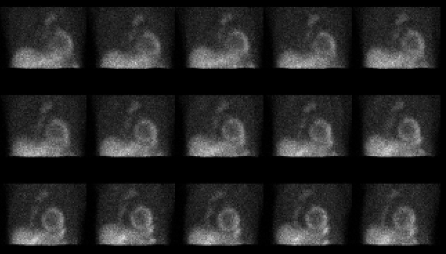
Selected images from the raw projection data acquired during the exercise portion of the myocardial perfusion study.
View main image(mi) in a separate viewing box
View second image(ct). Axial image of chest from contrast-enhanced computed tomography
View third image(xr). Posteroanterior chest
Full history/Diagnosis is also available
Return to the Teaching File home page.