
After viewing the image(s), the Full history/Diagnosis is available by using the link here or at the bottom of this page
Vertical long axis (left)and quality control images(right); the quality control images consist of a sinogram and selective linogram.
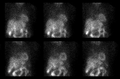
Top frames represent stress sestamibi images, and resting thallium imaging in lower frames
View main image(mi) in a separate viewing box
View second image(mi). Selected stress projection images are also available, if desired.
Full history/Diagnosis is also available
Return to the Teaching File home page.