
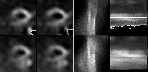
Vertical long axis (left)and quality control images(right); the quality control images consist of a sinogram and selective linogram.
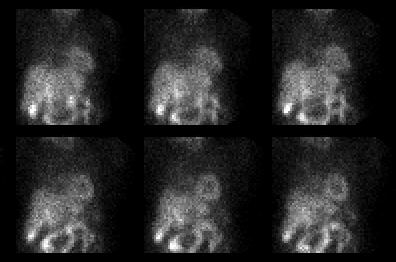
Top frames represent stress sestamibi images, and resting thallium imaging in lower frames
View main image(mi) in a separate image viewer
View second image(mi). Selected stress projection images are also available, if desired.
Full history/Diagnosis is available below
However, there is a "break" or discontinuity in the stress sinogram at the midpoint in the image, and there is moderate up/down motion of the cardiac activity (the horizontal white band) on the linogram. On review of the cine of the projection frames, these corresponded to sites of patient movement.
The repeat stress images show the perfusion to the inferior wall to be much better than on the initial stress images. The degree of decrease in the inferior wall is now the same on stress and rest images, and is most consistent with diaphragmatic attenuation.
View followup image(mi). Repeat stress images at 4 hours post initial injection, after re-emphasizing to the patient the importance of lying still during image acquisition.
Review of the projections for patient motion is critical, and can be done either by inspection of the projection data in cine format or by use of sinogram and linogram images.
The slow washout rate of Sestamibi permits reacquisition of the stress images within a few hours with minimal loss in image quality.
However, inspection of the projection images shown above demonstrate the liver to have a similar degree of activity as does the heart. If the "hot liver" artifact were present, the liver would typically have significantly more activity than the heart on anterior projection images.
References and General Discussion of Myocardial Imaging (Anatomic field:Heart and Great Vessels, Category:Other(Artifact))
Return to the Teaching File home page.