After viewing the image(s), the Full history/Diagnosis is available by using the link here or at the bottom of this page

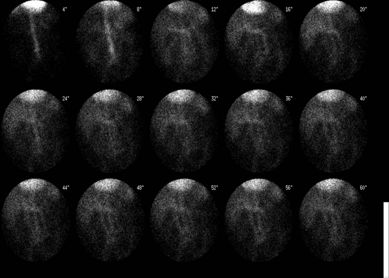
Initial flow images
View main image(gi) in a separate viewing box
View second image(gi). Delayed anterior projection images over 60 minutes
View AVI file of the entire acquisition (1.5 meg). (Note that the image has been set to higher intensity to reveal fainter portions of the image, resulting in pixel overflow in the cardiac region)
Full history/Diagnosis is also available
Return to the Teaching File home page.