Case Author(s): Akash Sharma, M.D. and Henry D. Royal, M.D. , 09/21/2004 . Rating: #D2, #Q4
Diagnosis: Damaged collimator artifact
Brief history:
88 year old patient admitted to the ICU with acute lower gastrointestinal bleeding.
Images:
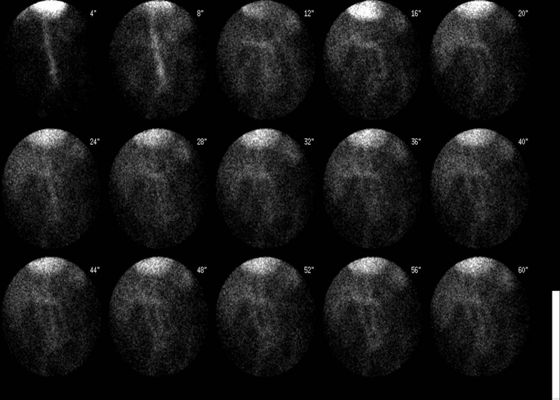
Initial flow images
View main image(gi) in a separate image viewer
View second image(gi).
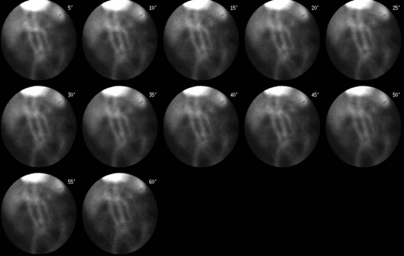
Delayed anterior projection images over 60 minutes
View AVI file of the entire acquisition (1.5 meg). (Note that the image has been set to higher intensity to reveal fainter portions of the image, resulting in pixel overflow in the cardiac region)
Full history/Diagnosis is available below
Diagnosis: Damaged collimator artifact
Full history:
88 year old patient admitted to the hospital with acute lower gastrointestinal bleeding. The patient has a history of left hemicolectomy in the past due to colon cancer. During this stay he had multiple episodes of bleeding per rectum and was transferred to the intensive care unit. A portable study was requested.
Radiopharmaceutical:
26.3 mCi Tc-99m in vitro labeled red cells i.v.
Findings:
No evidence for active gastrointestinal bleeding was seen on this study. However, on review of all static and cine images there is a persistent linear absence of activity projecting over the spleen. While the patient clearly moves on the cine projection, this linear focus remain immobile.
Discussion:
A linear area of decreased or absent activity raises the possibility of an artifact. Given that the area in question is fixed in location on a dynamic acquisition it is less likely to be on the patient's body (e.g. a line or a metallic photon attentuating object) and more likely to be camera related.
The two possibilities for a camera related artifact are intrinsic, such as a cracked cystal or extrinsis such as a damaged collimator.
Usually a cracked crystal defect also has characteristic 'edge-packing' presumed to be related to the refraction related to the photons incident on the edges of the crack. This is seen as a greater amount of activity on both sides of a photopenic defect (the crack itself with no photons registered).
In this case, the liner photopenic area does not have edge packing and is more likely to be related to the septa of the collimator, which have bent inward to nearly completely block any photons from striking the crystal surface in this region.
Followup:
A visual inspection of the collimator showed damage to the septa corresponding in length and location to the photopenic area on the image.
View followup image(mc).
Surface of the damaged collimator AFTER repair.
ACR Codes and Keywords:
References and General Discussion of Gastrointestinal Bleeding Scintigraphy (Anatomic field:Vascular and Lymphatic Systems, Category:Other(Artifact))
Search for similar cases.
Edit this case
Add comments about this case
Return to the Teaching File home page.
Case number: gi010
Copyright by Wash U MO