After viewing the image(s), the Full history/Diagnosis is available by using the link here or at the bottom of this page
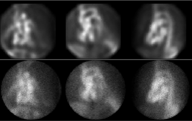
Radionuclide ventriculogram (filtered images above, unfiltered below). Note shape of inferior wall on lateral view. What is causing this contour?
View main image(ca) in a separate viewing box
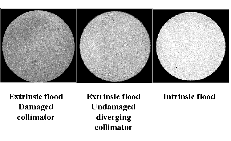
View second image(mm). Flood field
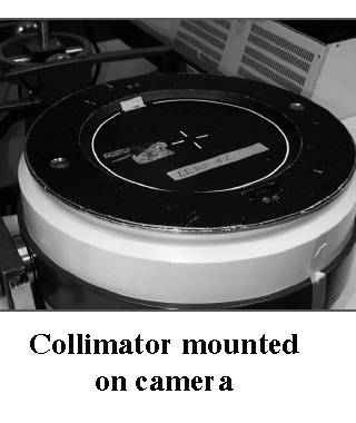
View third image(mm). Collimator
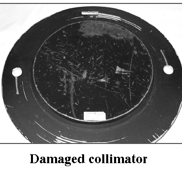
View fourth image(mm). Damaged collimator
Full history/Diagnosis is also available
Return to the Teaching File home page.