Case Author(s): David A. Hillier, M.D., Ph.D. and Jerold Wallis, M.D. , . Rating: #D2, #Q4
Diagnosis: Collimator damage
Brief history:
60 yo man with COPD undergoing lung transplant evaluation.
Images:
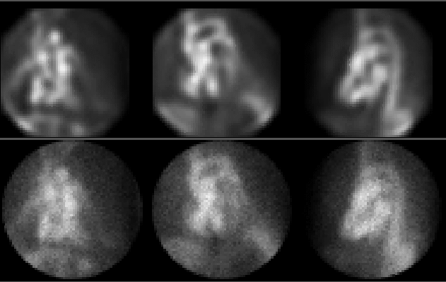
Radionuclide ventriculogram (filtered images above, unfiltered below).
Note shape of inferior wall on lateral view. What is causing this
contour?
View main image(ca) in a separate image viewer
View second image(mm).
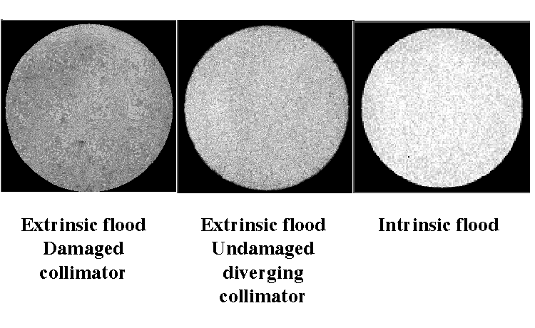
Flood field
View third image(mm).
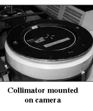
Collimator
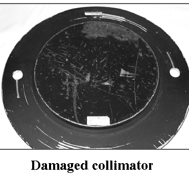
View fourth image(mm).
Damaged collimator
Full history/Diagnosis is available below
Diagnosis: Collimator damage
Full history:
60 yo man with COPD undergoing lung transplant evaluation.
Radiopharmaceutical:
26.1 mCi in-vivo labeled red cells
Findings:
Cardiac blood pool imaging, end systolic and end diastolic with and without filtering:
- Low-normal left ventricular function. LVEF = 55%
- Mildly reduced right ventricular function. RVEF = 32%
- Borderline bi-atrial enlargement.
- Defect apparently at inferior aspect of heart (especially on lateral view). Defect is at fixed position with respect to the camera.
2. Flood field:
- Extrinsic parallel hole collimator (used for clinical study above): Multiple small defects.
- Intrinsic flood: uniform.
- Extrinsic flood with diverging collimator: uniform.
Discussion:
The flood field for this camera is obtained with a point source (allowing only intrinsic flood fields to be obtained routinely, since the collimator septae would block the photons at the edges of the collimator). A sheet source would be necessary to perform an extrinsic (with collimator) flood field. This case shows the importance of performing extrinsic floods.
Collimators may be manufactured with septae constructed of lead film. Alternatively, they may be cast. The cast type are more durable. This collimator is of a cast design.
Differential Diagnosis List
A left ventricular defect is seen, particularly on the lateral view. The differential includes causes of filling defects, such as thrombus. However, the apparent defect is located at a constant location with respect to the camera. Artifact should be suspected. An extrinsic flood field (with collimator) was obtained with the collimator used in the study (parallel-hole collimator). This was obtained with a sheet source, revealing several small defects. An intrinsic flood field (without collimator) was normal. An extrinsic flood field on a different collimator (a diverging collimator) was normal. Visual inspection of the parallel-hole collimator revealed damage with dings and flattened areas in an identical pattern as that seen in the flood-field.
ACR Codes and Keywords:
References and General Discussion of Cardiac Blood Pool Scintigraphy (Anatomic field:Heart and Great Vessels, Category:Other(Artifact))
Search for similar cases.
Edit this case
Add comments about this case
Return to the Teaching File home page.
Case number: ca006
Copyright by Wash U MO