After viewing the image(s), the Full history/Diagosis is available by using the link here or at the bottom of this page

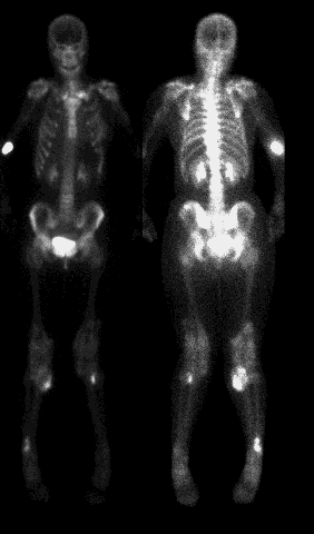
Anterior and posterior whole body images (the gray scale on the posterior image was intentionally changed to accentuate the findings)
View main image(bs) in a separate viewing box
View second image(xr). Anterior view of the right knee
View third image(xr). Lateral view of the right knee
Full history/Diagosis is also available
Return to the Teaching File home page.