Case Author(s): Charles Pringle, M.D./ Tom Miller, M.D. , 10/18/95 . Rating: #D2, #Q4
Diagnosis: Multiple hereditary exostoses
Brief history:
Increasing right leg pain.
Images:
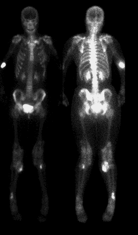
Anterior and posterior whole body images (the gray scale on the posterior image was intentionally changed to accentuate the findings)
View main image(bs) in a separate image viewer
View second image(xr).
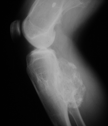
Anterior view of the right knee
View third image(xr).
Lateral view of the right knee
Full history/Diagnosis is available below
Diagnosis: Multiple hereditary exostoses
Full history:
16-year old female patient with
history of multiple hereditary exostoses and multiple
prior surgeries. Now, the patient has increasing right
leg pain.
Radiopharmaceutical:
19.5 mCi Tc-99m MDP
i.v.
Findings:
A focus of increased activity in the
medial right tibia corresponds to the symptomatic
area. There is also mildly increased activity about
both knees extending outside the normal boundaries
of the bones. Additional foci in the distal right and
proximal left tibio-fibular regions are also seen. Plain
radiographs of the right knee show multiple exostoses
with increase in size of the posterior tibial exostosis
since a previous study.
Discussion:
Multiple hereditary exostoses is
an autosomal dominant disorder. The lesions are
usually multiple and often bilateral, with the most
common sites being the knee, pelvis, rib, scapula and
elbow. The lesions generally stop growing when the
nearest epiphyseal center fuses. Malignant
transformation to chondrosarcoma occurs in less than
5% of cases.
Followup:
The symptomatic right tibial lesion
was surgically removed with pathologic findings
consistent with a benign osteochondroma. The other
two lesions seen on bone scintigraphy are being
followed clinically.
Major teaching point(s):
The increased activity in the
tibial lesion indicates increased metabolic activity but
is not specific for malignant degeneration.
ACR Codes and Keywords:
References and General Discussion of Bone Scintigraphy (Anatomic field:Skeletal System, Category:Normal, Technique, Congenital Anomaly)
Search for similar cases.
Edit this case
Add comments about this case
Read comments about this case
Return to the Teaching File home page.
Case number: bs036
Copyright by Wash U MO