After viewing the image(s), the Full history/Diagnosis is available by using the link here or at the bottom of this page
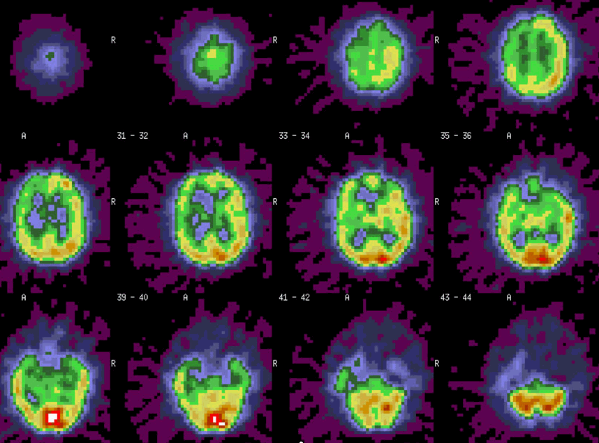
Initial brain SPECT with Technetium-99m-Bicisate intravenous injection during balloon occlusion of the right carotid artery. Imaging was performed one hour later.
View main image(br) in a separate viewing box
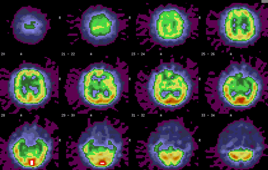
View second image(br). A brain SPECT was repeated the next day without right carotid artery occlusion to assess perfusion at baseline.
View third image(mr). MRI shows the cystic portion of the right skull base lesion adjacent to the cavernous portion of the right carotid artery.
Full history/Diagnosis is also available
Return to the Teaching File home page.