Case Author(s): Jabi Shriki, M.D., and Tom R. Miller, M.D., Ph.D. , 06/21/06 . Rating: #D2, #Q4
Diagnosis: Carotid occlusion study demonstrating insufficient contralateral flow for carotid artery sacrifice.
Brief history:
42-year-old man with a right skull base lesion.
Images:
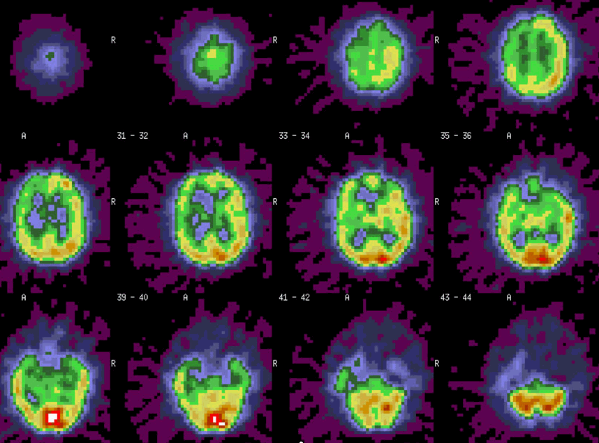
Initial brain SPECT with Technetium-99m-Bicisate intravenous injection during balloon occlusion of the right carotid artery. Imaging was performed one hour later.
View main image(br) in a separate image viewer
View second image(br).
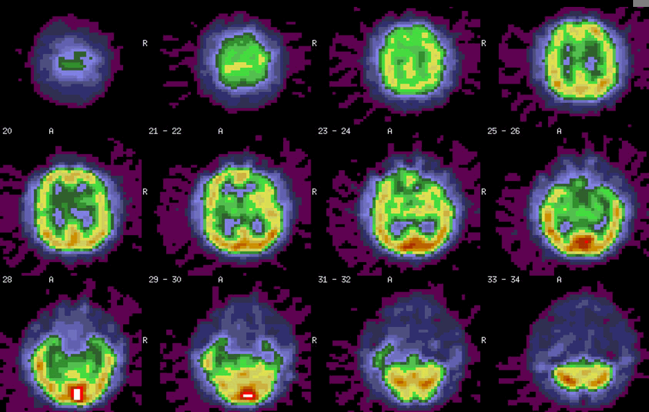
A brain SPECT was repeated the next day without right carotid artery occlusion to assess perfusion at baseline.
View third image(mr).
MRI shows the cystic portion of the right skull base lesion adjacent to the cavernous portion of the right carotid artery.
Full history/Diagnosis is available below
Diagnosis: Carotid occlusion study demonstrating insufficient contralateral flow for carotid artery sacrifice.
Full history:
This is a 42-year-old man with a right skull base lesion. Slow growth was demonstrated on serial radiographic studies. Multiple prior biopsy specimens yielded only skeletal muscle. More definitive excisional biopsy was planned, but it was felt that there was sufficient involvement of the right carotid artery that the artery might have to be sacrificed during the biopsy.
Radiopharmaceutical:
Technetium-99m-Bicisate i.v.
Findings:
SPECT images of the brain during right internal carotid artery balloon occlusion (main images) and at baseline (second images) demonstrate the decrement in perfusion of the right hemisphere of the brain during occlusion. This suggested that collateral flow provided from the left internal carotid artery alone might be insufficent to prevent infarction if the right carotid was sacrificed during biopsy.
Discussion:
Tc99m-Bicisate (L,L-Ethyl Cysteinate Dimer (L,L-ECD))and Tc99m-HMPAO (Hexamethylpropyleneamine Oxime) are also known as Neurolite and Ceretec. Both are lipophilic cations which cross the blood brain barrier. Initial uptake is proportional to blood flow, and there is minimal redistribution after uptake. Uses of technetium-based tracers for imaging the brain also include evaluation of patients with seizures, using ictal and interictal imaging.
References:
Van Paesschen W. Ictal SPECT. Epilepsia. 2004;45 Suppl 4:35-40.
Followup:
Further excisional biopsy was subsequently performed with sparing of the right internal carotid artery.
Major teaching point(s):
SPECT imaging during balloon occlusion can be helpful when carotid artery sacrifice is contemplated. Because there is a small normal variation in perfusion of the two hemispheres, a baseline study is often performed when occlusion imaging is asymmetric.
ACR Codes and Keywords:
References and General Discussion of Brain Scintigraphy (Anatomic field:Skull and Contents, Category:Neoplasm, Neoplastic-like condition)
Search for similar cases.
Edit this case
Add comments about this case
Return to the Teaching File home page.
Case number: br005
Copyright by Wash U MO