Case Author(s): Rusty Roberts, M.D. and Tom R. Miller, M.D., Ph.D. , . Rating: #D4, #Q4
Diagnosis: Occlusive pulmonary emobolism
Brief history:
82 year old woman with alpha-1-antitrypsin deficiency presents with shortness of breath.
Images:
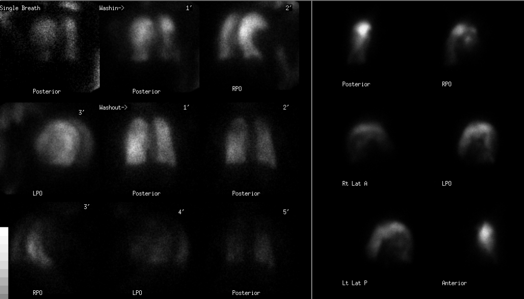
Xe-133 ventilation images and Tc-99m MAA perfusion images
View main image(vq) in a separate image viewer
View second image(xr).
PA and lateral chest radiographs
Full history/Diagnosis is available below
Diagnosis: Occlusive pulmonary emobolism
Full history:
82 year old woman with progressive shortness of breath presented to the emergency department with worsening dyspnea and tachypnea. She has known apha-1-antitrypsin deficiency.
Radiopharmaceutical:
28.3 mCi Xe-133 gas by inhalation and 4.2 mCi Tc-99m MAA i.v
Findings:
The Xe-133 ventilation images show bilateral hypoventilation of the mid and lower lung fields, with relative sparing of the upper lobes on single-breath and washin images. There is significant Xe-133 retention within the mid and lower lung fields bilaterally on the washout images.
The perfusion images demonstrate complete absence of radiotracer within the right lung. There is a suggestion of moderate-sized perfusion defects in the apicoposterior and anterior segments of the left upper lobe. There is hypoperfusion of the mid and lower lung fields in the left lung. This hypoperfusion matches the retention on the ventilation images.
The chest radiographs demonstrate a paucity of bronchovascular markings particularly within the lower lobes bilaterally consistent with emphysematous changes due to patient's known Alpha -I antitrypsin deficiency. There is relative opacification of the upper lobes bilaterally, left greater than right which may represent sparing of the upper lobe lung parenchyma versus a focal infiltrate within the upper lobes bilaterally, left greater than right. No pneumothorax or pleural effusion is seen. The aorta is mildly tortuous. The cardiac size is within normal limits. Diffuse osteopenia is noted.
Discussion:
This is an unusual presentation of pulmonary embolism because of the unilateral absence of perfusion to the right lung. However, this is very worrisome for pulmonary embolism because of the left upper lobe perfusion abnormalities. Pulmonary emobolii generally affect more peripheral pulmonary arteries even when a "saddle" embolus occurs. This distal embolization will result in the classic wedge defects. Rarely is an entire lung absent as in this case.
Followup:
A CT with performed using a pulmonary embolism protocol.
View followup image(ct).
Axial CT images with contrast in pulmonary arteries are shown. The top row is more cranial than the bottom row.
Differential Diagnosis List
The differential diagnosis includes pulmonary embolism, an obstructing mediastinal mass, mediastinal fibrosis, Swyer-James syndrome, pulmonary agenesis, pulmonary artery hypoplasia or agenesis and single pneumonectomy.
Datz, Fredrick L. Gamuts in Nuclear Medicine. Salt Lake City, Utah. Mosby 3rd ed., p 188, 1995.
ACR Codes and Keywords:
References and General Discussion of Ventilation Perfusion Scintigraphy (Anatomic field:Lung, Mediastinum, and Pleura, Category:Organ specific)
Search for similar cases.
Edit this case
Add comments about this case
Return to the Teaching File home page.
Case number: vq054
Copyright by Wash U MO