Case Author(s): Rusty Roberts, M.D. and Farrokh Dehdashti, M.D. , 11-12-04 . Rating: #D5, #Q5
Diagnosis: Fibrosing mediastinitis
Brief history:
40-year-old female with chest pain, cough, hemoptysis, and dyspnea. Noted to have lymphadenopathy (calcified) within the mediastinum on chest x-ray.
Images:

Xenon-133 ventilation and Tc-99m MAA perfusion images.
View main image(vq) in a separate image viewer
View second image(xr).
PA and lateral chest radiographs.
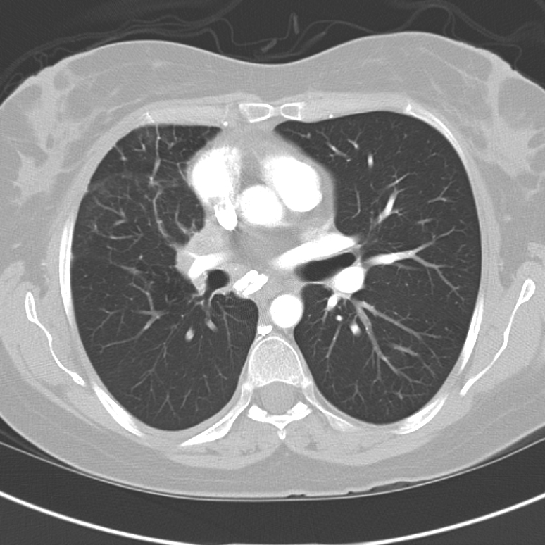
View third image(ct).
Axial CT image in pulmonary windows.
Full history/Diagnosis is available below
Diagnosis: Fibrosing mediastinitis
Full history:
40-year-old white female presented with no significant past medical history. She was transferred in from an outside hopital, secondary to "persistent pneumonia" and "abnormal chest computerized tomography." She started to notice shortness of breath three months prior to admission while she was climbing up stairs. She also felt chest tightness. However, she had no symptoms of chest pain, cough, fever or chills. She went to her primary care physician and was diagnosed community acquired pneumonia in 4 months prior to admission. She was given Levaquin and Rocephin. The symptoms resolved. Her symptoms returned and she went back to her primary care physician. She had a CT scan which showed right hilar lymphadenopathy with right middle lobe mild infiltration.
Radiopharmaceutical:
19.9 mCi Xe-133 gas by inhalation and 4.13 mCi Tc-99m MAA i.v.
Findings:
VQ: The Xe-133 images show a uniform distribution of activity on single breath and washin images. There is no abnormal Xe-133 retention during the washout phase. The perfusion images show absence of perfusion to the right lung. Perfusion of the left lung is physiologic with no perfusion defects.
CXR: Mild volume loss in the right lung is present. There is minimal increased opacity in the right hilum suggestive of lymphadenopathy. No pneumothorax is seen. The heart size is normal. No pleural effusion identified. Retained contrast is noted in the splenic flexure of the colon.
CT: Narrowed right pulmonary artery as is passes through a soft tissue attenuation mass along the right mediastinum with some calcifications. Mild ground glass attenuation in the right middle lobe.
Discussion:
Fibrosing mediastinitis in the United States is rare and usually idiopathic, however, an abnormal reaction to Histoplasma capsulatum may also be the cause in some patients. A proliferation of acellular collagen and fibrous tissue is seen within the pathologic samples.
"Affected patients are typically young and present with signs and symptoms of obstruction or compression of the superior vena cava, pulmonary veins or arteries, central airways, or esophagus. There may be two types of fibrosing mediastinitis: focal and diffuse. The focal type usually manifests on computed tomographic (CT) or magnetic resonance (MR) images as a localized, calcified mass in the paratracheal or subcarinal regions of the mediastinum or in the pulmonary hila. The diffuse type manifests on CT or MR images as a diffusely infiltrating, often noncalcified mass that affects multiple mediastinal compartments. CT and MR imaging play a vital role in the diagnosis and management of fibrosing mediastinitis."
Treatments vary from local excision to antibiotics and steriods depending on patient, symptoms, and suspected etiology.
Rossi, S. E., et al. AFIP Archives: Fibrosing Mediastinitis, Radiographics. 2001;21:737-757.
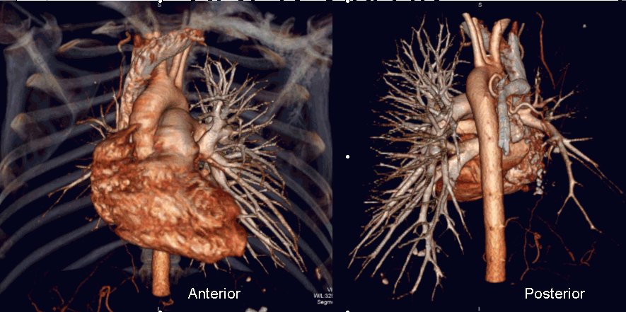
View followup image(ct).
CT pulmonary angiography reconstructed using a Vitrea 2 workstation.
Differential Diagnosis List
Obstructing mediastinal mass, lymphoma, fibrosing mediastinitis, or less likely a central pulmonary embolism.
ACR Codes and Keywords:
References and General Discussion of Ventilation Perfusion Scintigraphy (Anatomic field:Lung, Mediastinum, and Pleura, Category:Inflammation,Infection)
Search for similar cases.
Edit this case
Add comments about this case
Return to the Teaching File home page.
Case number: vq053
Copyright by Wash U MO