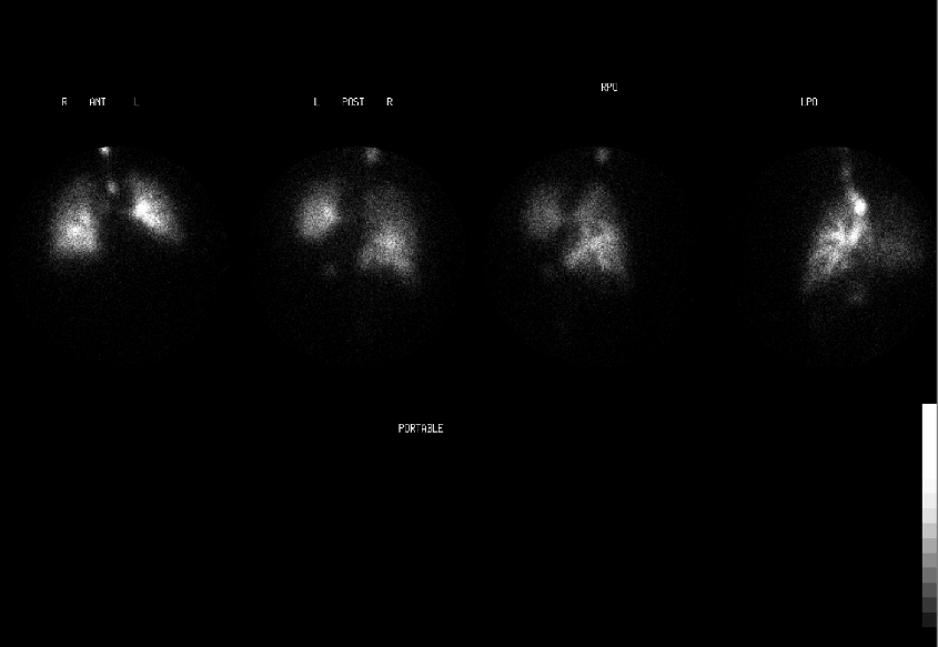
Ventilation images (anterior, posterior, RPO and LPO projections)
View main image(vq) in a separate image viewer
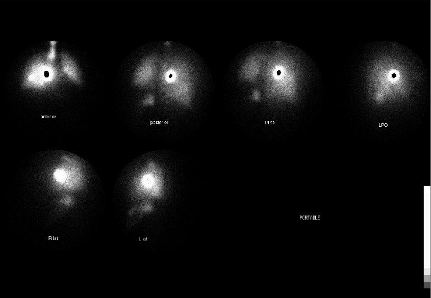
View second image(vq). Perfusion images (anterior, posterior, RPO, LPO projections, right lateral and left lateral projections)
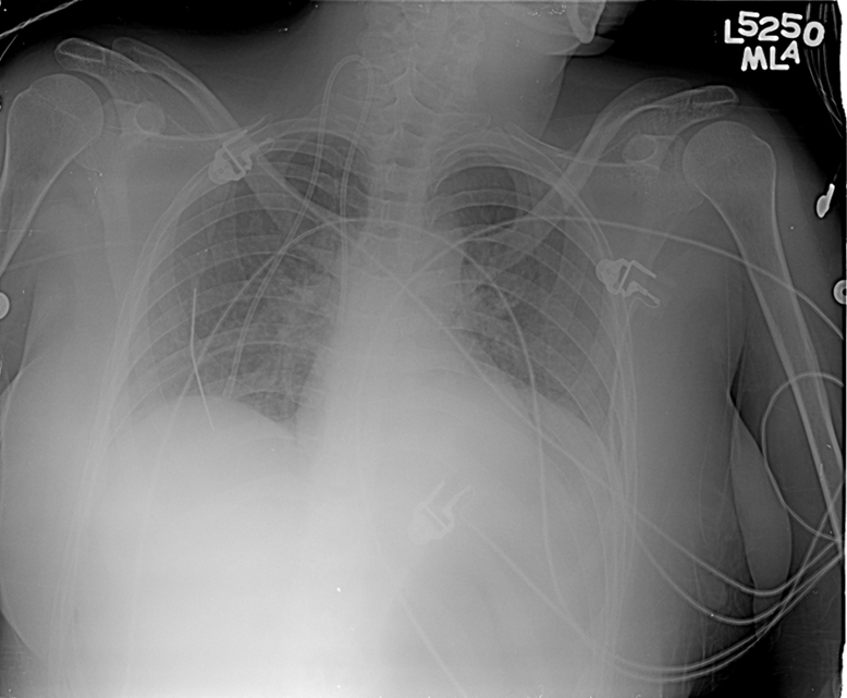
View third image(xr). Portable AP chest radiograph
View fourth image(vq). Re-windowed Perfusion images (anterior, posterior, RPO, LPO, right lateral and left lateral projections)
Full history/Diagnosis is available below
Due to her chronic medical conditions, she has a portacatheter present for IV access. For the perfusion examination, the patient was injected via the portacatheter.
Perfusion images: (rewindowed) Non diagnostic pulmonary perfusion examination due to lack of significant activity reaching the pre-capillary arterioles. The activity in the lungs seen on prior "perfusion" images was left over from the aerosol ventilation study.
Chest radiograph: Low lung volumes with normal cardiomediastinal shilouette. There are perihilar interstitial infiltrates and small bilateral pleural effusions. A right internal jugular central venous catheter line terminates at the superior vena.
In this case, nearly all of the radiolabelled MAA remained in the right upper chest (i.e. very little, if any, Tc99m MAA reached the pulmonary precapillary arterioles). Thus, the initial series of images labeled "pulmonary perfusion images" (image 2) shown actually represent residual activity from the ventilation examination. The images have set at a lower window threshold. This can be evidenced by comparing the intensity of tracer activity in structures such as the trachea and stomach on the ventilation (image 1) and "pulmonary perfusion images" (image 2). When the pulmonary perfusion images are re-windowed, the majority of radiopharmaceutical is noted in a single focus in the right upper chest. (image 4). Thus the perfusion examination is non-diagnostic.
Williams, SC (2001) Nuclear Medicine Online Reference Text. Ventilation/Perfusion Imaging: Technique & Radiopharmaceuticals. Retrieved February 10, 2004, from Aunt Minnie Web Site: http://www.auntminnie.com/default.asp?sec=ref&sub=ncm&pag=pul&itemid=54282
On review of image 2, the majority of radiolabelled MAA appears *within* the chest near the expected catheter tip rather than *on the chest wall*. Since fibrin sheaths/thrombi can form at the tips of chronic indwelling catheters, it was presumed that the activity was trapped in one of these sheaths, even though this would be quite unusual.
Because of the lack of activity reaching the pulmonary precapillary arterioles, the study was non diagnostic for the evaluation for pulmonary emboli. The examination was not repeated at the request of the referring clinicians. The patient expired from complications relating to her intracranial hemorrhage without further study of the pulmonary vasculature (angiography) or repeat ventilatory perfusion scintigraphy.
1) thrombus of activity formed from aspiration of blood into the syringe containing the radiolabelled MAA, and deposited in the lungs or adhering to the catheter tip.
2) activity remaining in the reservoir of a portacatheter, in the chest wall.
3) activity deposited on the skin surface, due to leakage during injection.
4) post-mortum injection of tracer through catheter, after cardiac circulation had ceased.
References and General Discussion of Ventilation Perfusion Scintigraphy (Anatomic field:Lung, Mediastinum, and Pleura, Category:Other(Artifact))
Return to the Teaching File home page.