Case Author(s): Jeffrey Yu, MD and Tom R. Miller, MD, PhD , 6/30/2000 . Rating: #D3, #Q5
Diagnosis: Brachiocephalic Vein Stenosis
Brief history:
45 year-old female with lupus presents with shortness of breath. Please rule out pulmonary embolism
Images:
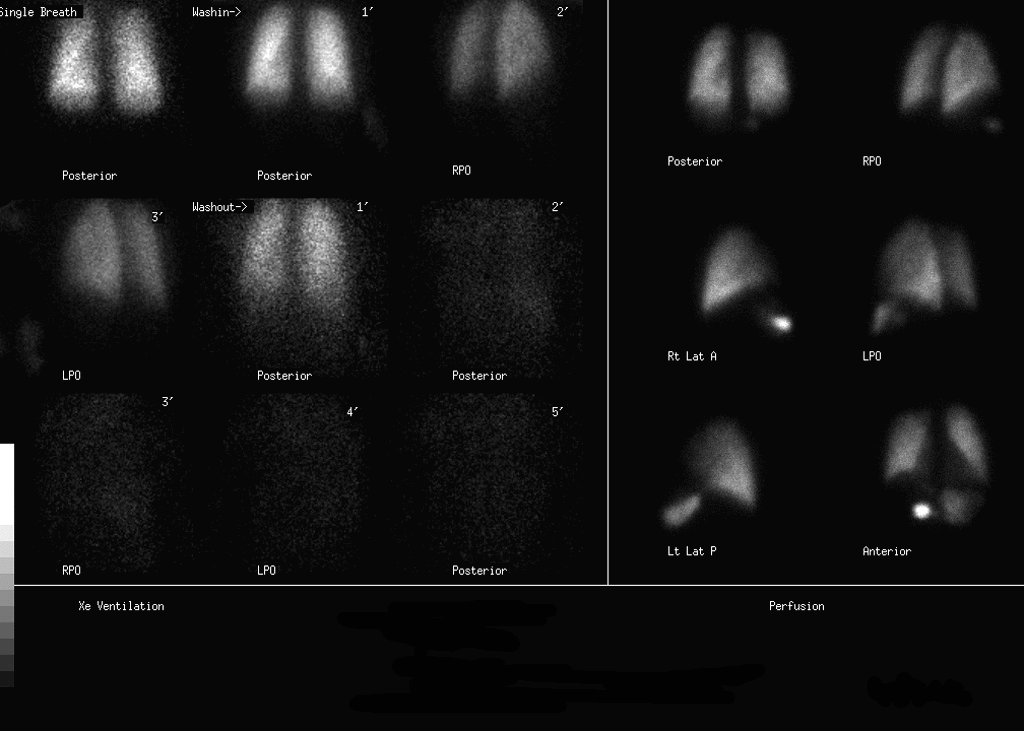
Dynamic ventilation and perfusion images in multiple projections
View main image(vq) in a separate image viewer
View second image(vq).
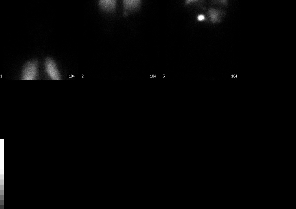
Spot images of the brain and abdomen
View third image(mc).
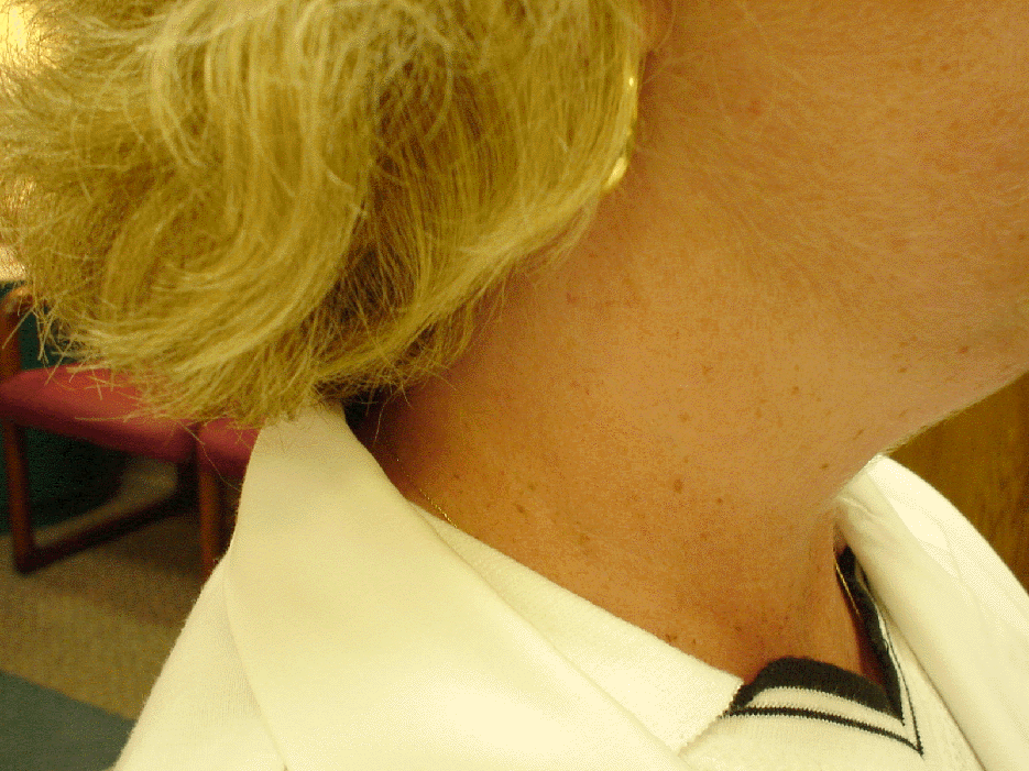
Picture of the patient's neck prior to bending over
View fourth image(mc).
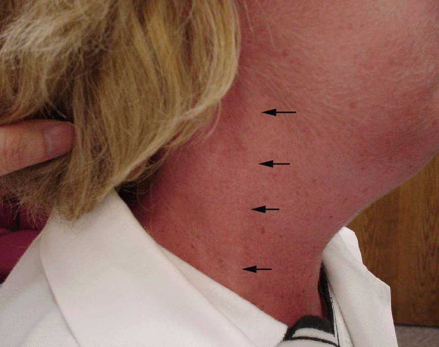
Picture of the patient's neck after she bent over with her head below her heart for a few seconds. Image exposure is unchanged relative to the previous image.
Full history/Diagnosis is available below
Diagnosis: Brachiocephalic Vein Stenosis
Full history:
45 year-old female with systemic lupus erythematosis who presents with increasing shortness of breath. We were asked to evaluate for pulmonary embolism. She has a history of prior pulmonary embolism in 1979 treated with heparin. She does not have an inferior vena filter caval filter.
Radiopharmaceutical:
Xe-133 and Tc-99m MAA
Findings:
The comparison chest radiograph performed on 6-22-00 demonstrates no pulmonary infiltrates or pleural fluid. The Xe-133 ventilation images show a small area of decreased activity on the single-breath image in the right lung apex. The washin images are unremarkable. There is no significant Xe-133 retention during the washout phase. There is a moderate-sized area of mildly decreased perfusion in the right lung apex matching the ventilatory abnormality. Otherwise, no unmatched perfusion defects are identified.
Of note, on the perfusion images there is a focus of increased activity likely in the quadrate lobe of the liver. In addition, there is activity in the left lobe of the liver.
Discussion:
Findings highly suggestive of partial superior vena caval obstruction. By history, the patient's porta catheter is still patent. However, the constellation of clinical findings as well as the appearance of a hot quadrate lobe and activity in the left lobe of the liver on ventilation-perfusion scintigraphy suggest superior vena caval obstruction.
Upon further questioning, the patient has been having difficulty with nausea and vomiting. When she bends over to vomit, she has noted persistent swelling in her neck. This was demonstrated to us in the Nuclear Medicine Department where she had persistent jugulo-venous distention as well as facial and neck plethora after bending over. In addition, she has a left subclavian porta catheter in place for administration of Cytoxan for lupus nephritis.
Followup:
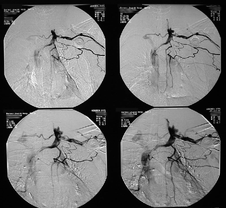
The scout radiograph demonstrates a left upper chest port with the catheter tip in the region of the right atrium.
With contrast injection, the left subclavian vein is somewhat small and contains a small linear filling defect. The innominate vein at its junction with the left subclavian vein is normal in caliber but then tapers down centrally before entering the superior vena cava. The superior vena cava appears normal in caliber and no thrombus is seen.
Multiple collateral vessels are seen in and around the left shoulder girdle with collaterals extending across the lower neck and perivertebral region to bypass the narrowing.
OPINION:
1. Chronic thrombi and narrowing involving the left subclavian vein and central left brachiocephalic vein as described above. Multiple collateral vessels are seen entering the left shoulder and chest and bypass these areas.
2. Normal appearing superior vena cava.
View followup image(an).
Left upper extremity venogram.
ACR Codes and Keywords:
References and General Discussion of Ventilation Perfusion Scintigraphy (Anatomic field:Lung, Mediastinum, and Pleura, Category:Other(Artifact))
Search for similar cases.
Edit this case
Add comments about this case
Read comments about this case
Return to the Teaching File home page.
Case number: vq045
Copyright by Wash U MO