Case Author(s): Daniel Apelbaum, MD and Henry Royal, MD , 5/8/00 . Rating: #D3, #Q5
Diagnosis: lymphangitic spread of tumor
Brief history:
41 year old female with respiratory distress.
Images:

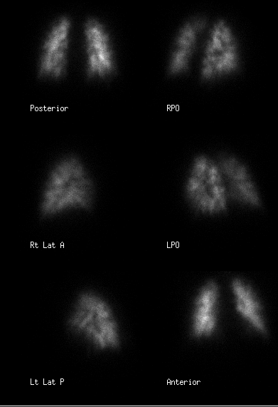
Images as labelled above.
View main image(vq) in a separate image viewer
View second image(vq).
Images as labelled above.
View third image(xr).
Current CXR.
View fourth image(ct).
Concurrent selected thoracic CT images.
Full history/Diagnosis is available below
Diagnosis: lymphangitic spread of tumor
Full history:
This is a 41 year old female with a history of metastatic cervical carcinoma. She became severely dyspneic and ventilation-perfusion scintigraphy was requested to evaluate for pulmonary embolism.
Radiopharmaceutical:
14.8 mCi Xe-133 gas via inhalation; 4.3 mCi Tc-99m MAA iv.
Findings:
The chest radiograph demonstrates the lungs to be essentially clear without detectable infiltrate or pleural fluid.
The ventilation images are normal. The perfusion images demonstrate the "segmental contouring" sign: perfusion defects are present at the very periphery of most segments.
On thoracic CT, there is no evidence of pulmonary embolism. However, nodular thickening of many of the interlobar septae is present, suggestive of lymphangitic spread of carcinoma. There are small pleural effusions. Mediastinal and hilar adenopathy is also present.
Discussion:
The segmental contouring sign indicates occlusion at the very small pulmonary arteriolar level. Instead of occlusion of the main, segmental, and/or medium-to-large subsegmental pulmonary arteries as is typically seen with embolism from venous thrombus, segmental contouring reflects a more distal occlusive process. A classical etiology is hematogenous microembolism from tumor (the precursor to lymphangitic spread), though other forms of microembolism, including fat and talc can cause this appearance. Any microvascular insult such as vasculitis or even sarcoidosis may also result in the segmental contour pattern.
ACR Codes and Keywords:
References and General Discussion of Ventilation Perfusion Scintigraphy (Anatomic field:Lung, Mediastinum, and Pleura, Category:Neoplasm, Neoplastic-like condition)
Search for similar cases.
Edit this case
Add comments about this case
Return to the Teaching File home page.
Case number: vq044
Copyright by Wash U MO

