Case Author(s): Lester Johnson, M.D., Ph.D. and Jerold Wallis, M.D. , 1/14/00 . Rating: #D4, #Q4
Diagnosis: Takayasu arteritis
Brief history:
Pleuritic chest pain and dyspnea, rule out pulmonary embolism
Images:

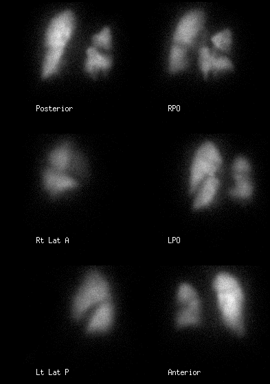
Perfusion images
View main image(vq) in a separate image viewer
View second image(vq).
Ventilation images
View third image(xr).
A PA chest radiograph is shown
View fourth image(mr).
A coronal oblique image from pulmonary magnetic resonance angiography is shown
Full history/Diagnosis is available below
Diagnosis: Takayasu arteritis
Full history:
This 42-year-old woman has a history of Takayasu arteritis. The perfusion scan abnormalities are unchanged in comparison to a previous V/Q scan obtained 8 years earlier (not shown). Pulmonary angiography performed in the past demonstrated findings consistent with progressive Takayasu arteritis (not shown).
Radiopharmaceutical:
Xe-133 gas by inhalation and Tc-99m macroaggregated albumin (MAA) by intravenous injection
Findings:
The comparison chest radiograph demonstrates small
bilateral pleural effusions, right greater than left. There
is a small amount of atelectasis or infiltrate at the left
lung base. The Xe-133 ventilation images show a
uniform distribution of activity on single-breath and
washin images. There is no abnormal Xe-133
retention during the washout phase. The perfusion
images show decreased activity within the superior
and posterobasal segments of the left lower lobe.
There is also decreased perfusion within the lingula. The
right lung demonstrates large segmental defects in all
lobes, unchanged in comparison to the previous
examination.
Magnetic resonance images of the chest demonstrate
multiple areas of stenosis of the great vessels
(brachiocephalic and left internal carotid), without
significant change since seven months earlier. Multiple
areas of stenosis and occlusion were also noted in
both left and right pulmonary arterial systems (not
shown), also unchanged.
Discussion:
Arteritis which involves medium to large vessels, such as polyarteritis nodosa and Takayasu arteritis, can cause mismatched subsegmental perfusion defects and hence are a known potential mimic of pulmonary embolism on ventilation/perfusion scintigraphy. The clinical history and comparison to prior studies are crucial for correctly interpreting these studies. Other potential mimics of acute pulmonary embolism include idiopathic pulmonary fibrosis (IPF) and radiation pneumonitis (which may pathologically be a type of vasculitis), generally seen in elderly patients, and multiple peripheral pulmonic stenoses, a congenital abnormality generally diagnosed in childhood. It should be noted that these moderate to large segmental perfusion defects are usually distinct from the small defects (sometimes referred to as "ratbites") that may be seen with small vessel vasculitis, such as Lupus, or pulmonary hypertension.
ACR Codes and Keywords:
References and General Discussion of Ventilation Perfusion Scintigraphy (Anatomic field:Vascular and Lymphatic Systems, Category:Other generalized systemic disorder)
Search for similar cases.
Edit this case
Add comments about this case
Return to the Teaching File home page.
Case number: vq043
Copyright by Wash U MO

