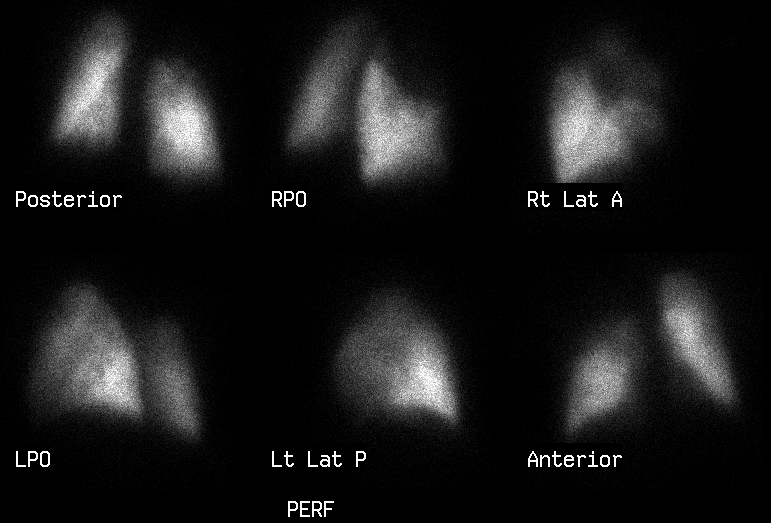

Initial pulmonary perfusion scintigrams performed on 7/16/99
View main image(vq) in a separate image viewer
View second image(vq). Subsequent Xe-133 ventilation images obtained on 8/25/99. Patient now has increased shortness of breath.
View third image(vq). Pulmonary perfusion images from 8/25/99
View fourth image(xr). Chest radiograph from 8/25/99.
Full history/Diagnosis is available below
The perfusion images show absent perfusion of the right upper lobe. Mild diffuse heterogeneity of perfusion is seen in the remainder of the right lung and throughout the left lung.
Ventilation-Perfusion Scintigraphy 8/25/99:
The patient returned for ventilation imaging to complement his earlier perfusion studyof 7/16/99. At this time, he was having increased shortness of breath.
The comparison chest radiograph demonstrates a large spiculateds right suprahilar mass. There is volume loss of the right upper lobe, with associated elevation of the right hemidiaphragm and shift of the mediastinum to the right. The Xe-133 washin ventilation images show almost no ventilation to the entire right lung. There is abnormal Xe-133 retention during the washout phase in the entire right lung.
Because of the markedly abnormal ventilation study, a repeat perfusion study was obtained. The perfusion images show almost no perfusion to the entire right lung. This is a marked change from the prior examination dated 7-16-99, when the right middle lobe and right lower lobe were well perfused and only the right upper lobe demonstrated very poor perfusion.
The near total lack of ventilation to the right lung on the 8/25/99 study suggests new involvement of the bronchus intermedius supplying the right middle and lower lobes (or central extension to involve the right main bronchus). The apparent mismatch of the findings on the ventilation study on this date with the perfusion study performed on 7/16/99 prompted a repeat perfusion study.
The new marked reduction in pulmonary blood flow to the right middle and lower lobes evident on the perfusion scan of 8/25/99 might make one suspect involvement of the pulmonary arteries (or veins) supplying these lobes. However, an outside hospital contrast-enhanced CT scan performed 9 days earlier on 8/16/99 demonstrated patent pulmonary arteries, thus contradicting this hypothesis and suggesing that the decrease in perfusion is due to auto-regulation (hypoxic vasoconstriction) of pulmonary blood flow secondary to decreased ventilation.
This study was obtained after therapeutic "shelling out" of tumor from the right middle and right lower lobe bronchi during bronchoscopy. This reveals marked improvement in ventilation and perfusion to the right middle and right lower lobes since 8-25-99. This proves that the marked decrease in perfusion is related to the poor ventilation caused by the endobronchial component of the patient's squamous cell cancer.
The post-procedure perfusion defect in the right upper lobe is similar to that seen on the original perfusion study of 7/16/99 and represents the effects of the residual endobronchial squamous cell carcinoma.
View followup image(vq). Post-bronchoscopic ventilation-perfusion images 8/30/99.
2) Temporal separation of ventilation and perfusion imaging can be misleading, and if there is any doubt, obtaining a contemporaneous perfusion or ventilation study is recommended.
2) Primary involvement of both the bronchus and pulmonary arteries/veins due to tumor extension.
References and General Discussion of Ventilation Perfusion Scintigraphy (Anatomic field:Lung, Mediastinum, and Pleura, Category:Neoplasm, Neoplastic-like condition)
Return to the Teaching File home page.