Case Author(s): Marc Montella, M.D. and Keith Fischer, M.D. , 09/27/99 . Rating: #D3, #Q3
Diagnosis: Intrapulmonary Physiologic Shunt
Brief history:
57 year old male in ICU with hypoxia.
Images:
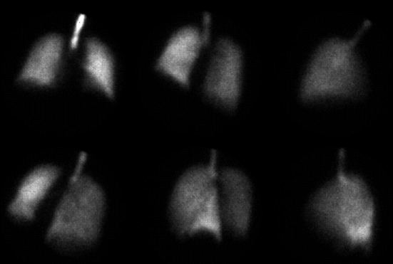
Tc-99m MAA Perfusion Images
View main image(vq) in a separate image viewer
View second image(vq).
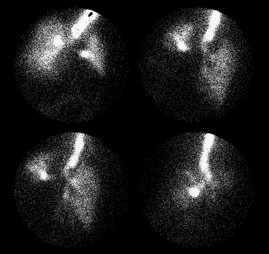
Tc-99m DTPA Aerosol Images (Ant, Post, RPO, LPO)
View third image(xr).
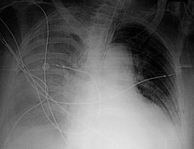
Portable AP Chest X-Ray
Full history/Diagnosis is available below
Diagnosis: Intrapulmonary Physiologic Shunt
Full history:
57 year old man with large left pleural effusion and a
questionable bronchus "cut-off" sign seen on standard chest radiograph. The patient had a desaturation event earlier today and the following examination is to evaluate for pulmonary embolism.
Radiopharmaceutical:
Tc-99m DTPA Aerosol and Tc-99m MAA
Findings:
The comparison chest radiograph demonstrates right
pleural effusion and a retrocardiac opacity compatible with
partial left lower lobe atelectasis. There is a smaller left
pleural effusion (post-thoracentesis).
The Tc-99m DTPA aerosol images demonstrate decreased ventilation,
with nearly absent ventilation to the left lower lobe and lingula.
The perfusion images demonstrate no segmental defects. There is,
however, increased relative perfusion to the left lower lobe as compared to the left upper lobe and entire right lung. The left lower lobe shows decreased volume consistent with atelectasis.
Discussion:
The normal vasoconstrictive response to hypoxia would cause shunting of perfusion away from hypoxic areas of the lung. When this does not occur as in this case, the result is hypoxemia produced by this physiologic shunt i.e. lung with little or no ventilation is still being perfused.
ACR Codes and Keywords:
References and General Discussion of Ventilation Perfusion Scintigraphy (Anatomic field:Lung, Mediastinum, and Pleura, Category:Misc)
Search for similar cases.
Edit this case
Add comments about this case
Return to the Teaching File home page.
Case number: vq040
Copyright by Wash U MO