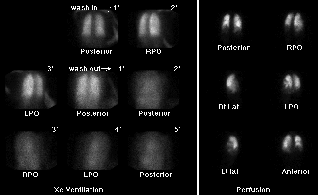
Xenon ventilation and perfusion images
View main image(vq) in a separate image viewer
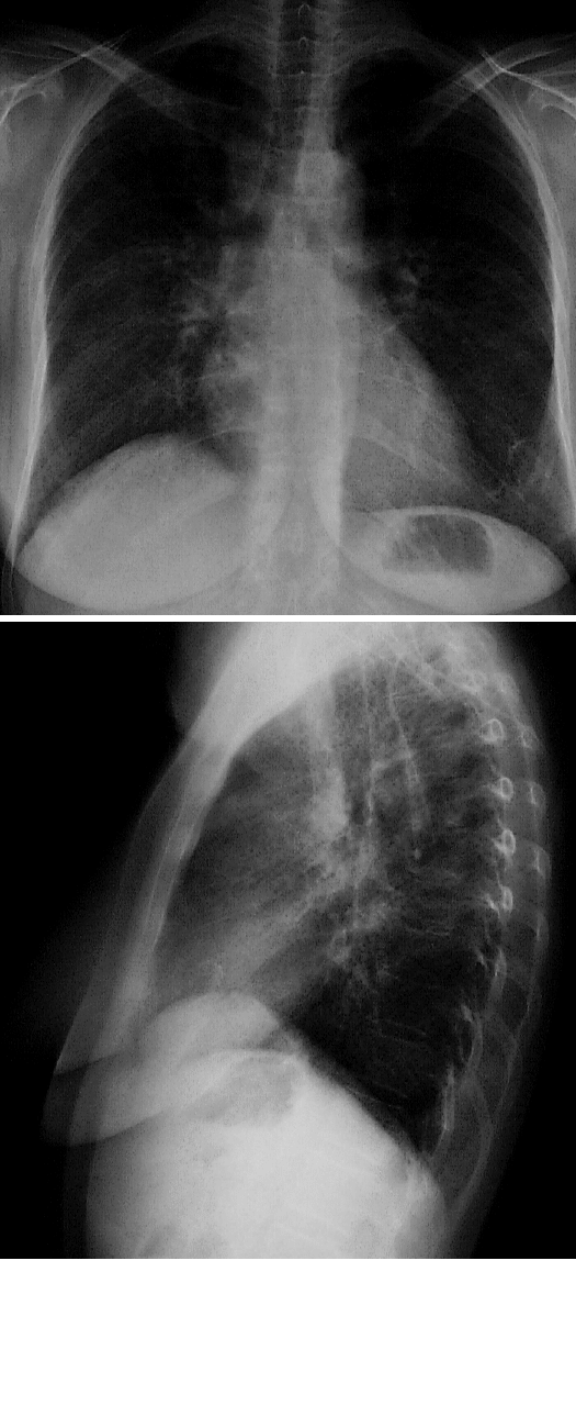
View second image(xr). PA and lateral chest radiograph
Full history/Diagnosis is available below
A comparison chest radiograph shows a pattern of reticular interstitial opacities in both lungs.
Studies done to assess the mechanism by which IPF causes this scintigraphic appearance have shown that the perfusion defects correspond to areas of cystic air spaces in the periphery of the lung in IPF patients. Unlike emphysema, these cystic spaces in IPF are ventilated normally, and therefore do not produce ventilatory defects which correspond to the perfusion abnormalities. In this case, the patient was without acute symptoms, and the study was done as part of a lung transplant evaluation. Assesssment for pulmonary embolism superimposed on a patient with IPF would be more problematic. Chest CT could be obtained to correlate the defects to areas of cystic spaces. Alternatively, the patient could be evaluated with pulmonary angiography or spiral CT to look for pulmonary embolism (though little data on spiral CT is available in this disorder).
Reference:
Strickland NH. Cause of regional ventilation perfusion mismatching in patients with idiopathic pulmonary fibrosis. AJR 1993; 16: 719-725
References and General Discussion of Ventilation Perfusion Scintigraphy (Anatomic field:Lung, Mediastinum, and Pleura, Category:Organ specific)
Return to the Teaching File home page.