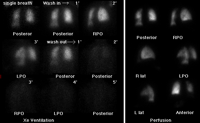
Xe-133 ventilation and Tc-99m MAA perfusion images.
View main image(vq) in a separate image viewer
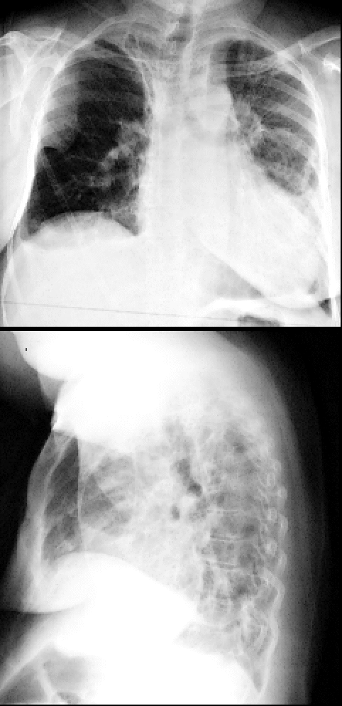
View second image(xr). PA and lateral chest radiograph
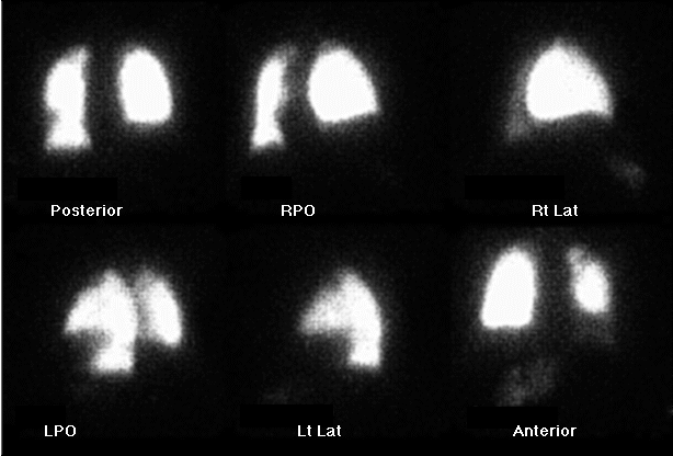
View third image(pe). Perfusion images windowed at higher intensity.
Full history/Diagnosis is available below
Perfusion images demonstrate a similar distribution of decreased activity in the left apex and left lower lobe. Subtle increased activity is seen in the right upper quadrant of the abdomen on the anterior and right lateral images.
Chest radiograph shows scarring in the lung apices as well as blunting of the right costophrenic angle.
Repeat view of the perfusion images is windowed at a higher intensity level. The images confirm activity in the anterior right upper quadrant, in the location of the liver.
The incidental finding of liver activity is of greater interest in this case. Visualization of the liver is an uncommon finding on a lung perfusion scan. The etiology is most often related to inferior or superior vena cava obstruction. The latter was determined clinically to be the etiology in this case. Collateral pathways for chronic superior vena cava obstruction have been well documented. These include internal mammary and epigastric veins, azygous and hemiazygous system, superior and inferior intercostal veins, and the paravertebral plexus. The collateral flow can result in shunting of MAA to the liver in general or the quadrate lobe specifically, as happened in this case. Additional causes of liver activity on the perfusion scan include intracardiac and intrapulmonary shunts.
Reference:
Edeburn GF, et al. AJR 1985, 145: 273-274.
References and General Discussion of Ventilation Perfusion Scintigraphy (Anatomic field:Gasterointestinal System, Category:Other generalized systemic disorder)
Return to the Teaching File home page.