Case Author(s): Jeff Chesnut, D.O. and Jerold Wallis, M.D. , 5/22/99 . Rating: #D3, #Q3
Diagnosis: Bronchopleural fistula
Brief history:
70 year old female 2 weeks status-post lung volume reduction surgery for emphysema. She now has increasing dyspnea.
Images:

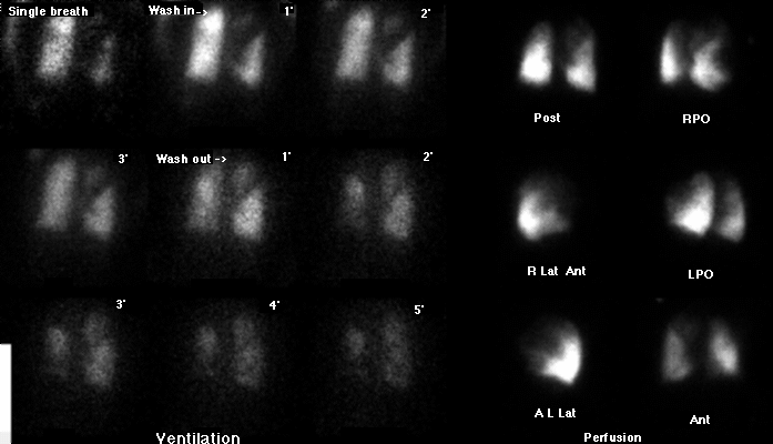
A ventilation/perfusion scintigram is shown. Does the
ventilation-perfusion mismatch at the left apex indicate a vascular
problem?
View main image(vq) in a separate image viewer
View second image(xr).
A portable chest radiograph is shown
(although difficult to see on this reproduction,there is a moderate
left apical pneumothorax with a chest tube in place).
Full history/Diagnosis is available below
Diagnosis: Bronchopleural fistula
Full history:
70 year old female 2 weeks status-post lung volume reduction surgery for emphysema. She now has increasing dyspnea.
Radiopharmaceutical:
Xe-131 gas and Tc-99m MAA
Findings:
The comparison chest radiograph demonstrates mild bilateral basilar infiltrates.
There is a small right sided pleural effusion. There is a moderate
left apical pneumothorax with a chest tube in place. (The chest radiograph
reproduced poorly. You'll have to trust me on this one.) The chest tube was on
suction during the examination.
The Xe-133 images show decreased activity at the right apex on the single-breath
and wash-in images. There is increased activity at the left apex in the
region of the pneumothorax. There is patchy diffuse retention of Xe-133
throughout both lungs on the wash-out views except in the left apex.
The perfusion images show a defect in the right lateral basilar segment in
the region of the pleural effusion. There are multiple areas of decreased
perfusion throughout which all match ventilatory abnormalities and are non-segmental.
Discussion:
The overall pattern of this study represents an intermediate liklihood ratio
for pulmonary embolus based on the right pleural effusion with matching
perfusion abnormality. The increased Xe-133 ventilation in the region of
the left apical pneumothorax represents a bronchopleural fistula.
Suction was on during the study with an active air leak, pulling xenon
into the pleural space and simulating well-ventilated lung on the
ventilation study.
ACR Codes and Keywords:
References and General Discussion of Ventilation Perfusion Scintigraphy (Anatomic field:Lung, Mediastinum, and Pleura, Category:Effect of Trauma)
Search for similar cases.
Edit this case
Add comments about this case
Return to the Teaching File home page.
Case number: vq036
Copyright by Wash U MO

