Case Author(s): Sarah Reimer, MD and Keith Fischer, MD , 04/23/99 . Rating: #D2, #Q3
Diagnosis: non-small cell lung cancer
Brief history:
54-year-old male with shortness of breath
Images:

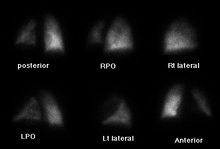
Perfusion scintigram
View main image(vq) in a separate image viewer
View second image(xr).
Ventilation scintigram
View third image(vq).
Portable chest radiograph
Full history/Diagnosis is available below
Diagnosis: non-small cell lung cancer
Full history:
This 54-year-old male presented to an outside hospital with shortness of breath. Ventilation perfusion scintigraphy performed there was interpreted as high likelihood ratio for pulmonary embolism. He was transferred to BJC, where repeat ventilation perfusion scintigraphy was performed. Lower extremity doppler examination was normal.
Radiopharmaceutical:
Xenon-133 via inhalation and Tc-99m MAA i.v.
Findings:
Ventilation images demonstrate diminished activity in the apices bilaterally on single-breath image with retention in the apices during the wash-out phase. The perfusion images demonstrate no perfusion to the left upper lobe and heterogeneous perfusion to the left lower lobe. Perfusion to the right lung is normal.
The portable chest radiograph demonstrates elevation of the left hemidiaphragm and a prominent left cardiac contour without infiltrate or pleural effusion.
Discussion:
Perfusion abnormalities which involve an entire lung or an entire lobe only should lower the suspicion of pulmonary embolism. Pulmonary embolism which could cause that extensive of an abnormality should be bilateral. Unilateral extensive perfusion abnormality is much more commonly seen with compression of a pulmonary artery by fibrosing mediastinitis or by a lung cancer or other tumor.
Followup:
CT was performed to look for fibrosing mediastinitis or lung tumor. Transbronchial biopsy of his middle mediastinal mass yielded non-small-cell lung cancer.
View followup image(ct).
CT of the chest demonstrates a middle mediastinal soft tissue mass which engulfs the vascular structures.
ACR Codes and Keywords:
References and General Discussion of Ventilation Perfusion Scintigraphy (Anatomic field:Lung, Mediastinum, and Pleura, Category:Neoplasm, Neoplastic-like condition)
Search for similar cases.
Edit this case
Add comments about this case
Read comments about this case
Return to the Teaching File home page.
Case number: vq034
Copyright by Wash U MO

