Case Author(s): Sarah Reimer, MD and Tom R. Miller, MD, PhD , 02/02/99 . Rating: #D4, #Q4
Diagnosis: tumor embolism
Brief history:
Pt. developed shortness of breath following a liver biopsy.
Images:

Ventilation images
View main image(vq) in a separate image viewer
View second image(vq).
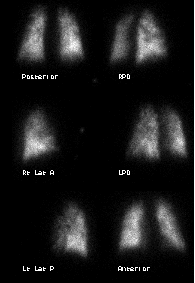
Perfusion images
Full history/Diagnosis is available below
Diagnosis: tumor embolism
Full history:
The patient is a 73-year-old male who had had an IVC filter placed five years prior to this study. An ultrasound examination demonstrated a liver mass, with biopsy revealing hepatocellular carcinoma. The patient experienced progressive shortness of breath around this time.
Findings:
The comparison chest radiograph (not shown) demonstrated hyperinflation without infiltrate or pleural effusion. The ventilation images demonstrate mild air trapping in the upper lobes. The perfusion images demonstrate multiple splinter-like, pleural based perfusion defects.
Discussion:
Metastatic tumor is an uncommon cause of pulmonary embolus, and the
diagnosis is usually made post-mortem. Diagnosis may be made pre-mortem by lung biopsy.
In contrast to embolic clot, which generally produces segmental defects on perfusion scintgraphy, embolic metastases generally produce very small, splinter-like peripheral defects. These may not be seen on pulmonary angiography. Pulmonary angiography or chest radiograph typically only show pulmonary artery enlargement due to severe pulmonary artery hypertension.
Followup:
The patient expired within days of this examination. Autopsy revealed diffuse tumor embolism throughout both lungs.
ACR Codes and Keywords:
References and General Discussion of Ventilation Perfusion Scintigraphy (Anatomic field:Lung, Mediastinum, and Pleura, Category:Neoplasm, Neoplastic-like condition)
Search for similar cases.
Edit this case
Add comments about this case
Return to the Teaching File home page.
Case number: vq033
Copyright by Wash U MO