Case Author(s): Matt Jaksha, M.D. and Farrokh Dehdashti, M.D. , . Rating: #D4, #Q4
Diagnosis: Vena cava obstruction and pulmonary embolism with right to left shunt
Brief history:
Rule out pulmonary embolism
Images:
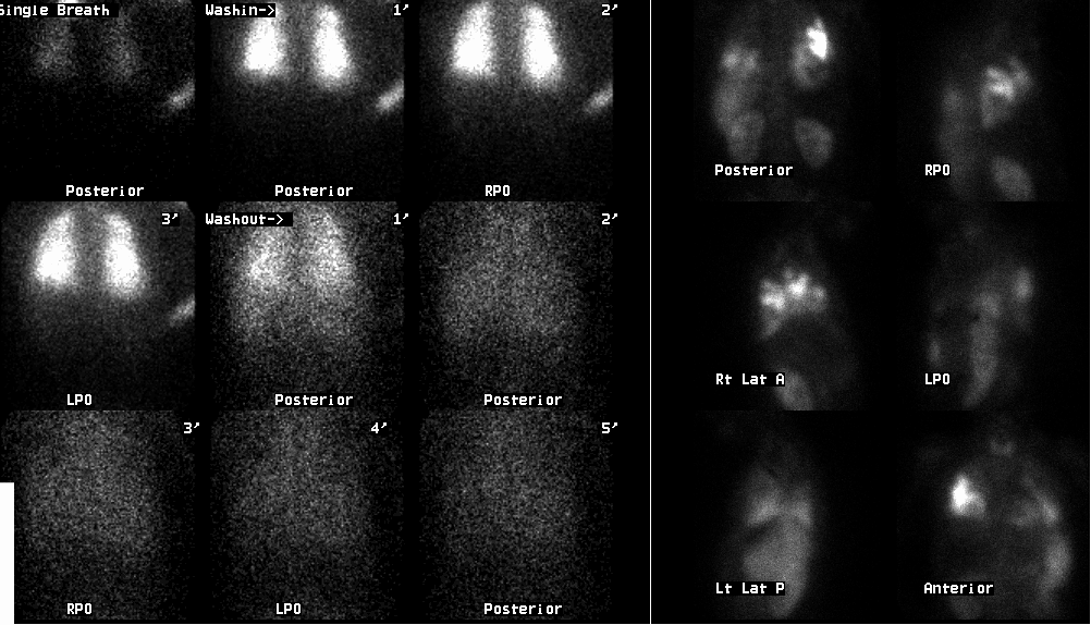
Ventilation and perfusion images
View main image(vq) in a separate image viewer
View second image(vq).
Posterior image of the abdomen and chest, posterior image of the head and neck
View third image(vq).
Pulmonary perfusion images from 5 years earlier
Full history/Diagnosis is available below
Diagnosis: Vena cava obstruction and pulmonary embolism with right to left shunt
Full history:
This is a 28 year old woman with a history of an autoimmune thrombotic disorder, and known thrombus involving the superior and inferior vena cava, as well as the portal system. She presents with marked hypoxia (pO2 = 42), and this study is requested to rule out pumonary embolism.
Radiopharmaceutical:
Xe-133 inhaled gas and Tc-99m macro-aggregated albumin
Findings:
The Xe-133 ventilation images show only mild heterogeneity. The perfusion
images show decreased perfusion to the majority of both lungs,
particularly the upper lungs. There are multiple wedge-shaped defects
bilaterally. Activity is seen in the kidneys and thyroid. Additional
images demonstrate significant activity within the brain. There is also
perfusion of a left upper quadrant organ, presumably the spleen.
The pulmonary perfusion study 5 years earlier showed normal perfusion of
the lungs, with activity seen in the upper abdomen anteriorly. At that
time, venography had demonstrated occlusion of the right subclavian vein
and superior vena cava, with multiple collateral vessels identified in
the body wall. There was no evidence of a right to left shunt. The
anterior abdominal activity represents particles in the left lobe of the
liver, predominantly the medial segment. This is commonly seen with SVC
occlusion and collateralization of body wall vessels, which drain to the
umbilical vein, through the liver and into the IVC.
Discussion:
While this woman had previously shown changes of SVC thrombosis, she had shown no evidence of pumonary embolus. Currently, there is evidence of extensive pulmonary emboli, or at least extensive thrombus in the pulmonary arteries, the origin of which may be complicated given her history. She has also developed extensive right to left shunting in the interval. The site of the shunt is not clear based on the scintigraphic images.
Followup:
The same day, the patient underwent a venous bubble examination to evaluate the level of the right to left shunt. Injection of bubbles into a right peripheral venous catheter showed flow directly into the left heart, bypassing the right heart. Subsequent venography showed tortuous chest wall collaterals which appeared to communicate with the intercostal veins and transpleurally communicate with the pumonary venous system.
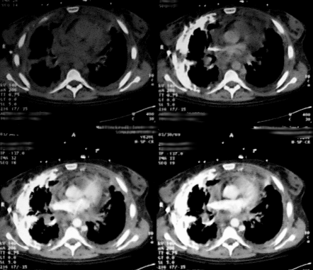
Finally, CT of the chest was performed dynamically at the level of the left atrium during contrast administration. The injection was made intravenously in the right forearm. The images demonstrate a large sheet of pleural collaterals enhancing in the right hemithorax, with subsequent enhancement of the left atrium. A large collateral vessel courses through the major fissure. The left atrium and aorta enhance long before the pulmonary artery. Note that the hemiazygous vein is markedly enlarged.
View followup image(ct).
Dynamic chest CT images with contrast injection in the right arm
ACR Codes and Keywords:
References and General Discussion of Ventilation Perfusion Scintigraphy (Anatomic field:Lung, Mediastinum, and Pleura, Category:Organ specific)
Search for similar cases.
Edit this case
Add comments about this case
Read comments about this case
Return to the Teaching File home page.
Case number: vq029
Copyright by Wash U MO