Case Author(s): Stephanie P.F. Yen, M.D. and Jerold Wallis, M.D. , 2/13/98 . Rating: #D3, #Q4
Diagnosis: Right-to-left Shunt Secondary to RUL Pulmonary Arteriovenous Malformation
Brief history:
67-year-old female who presents with shortness of breath, congestive
heart failure, and new onset atrial fibrillation.
Images:

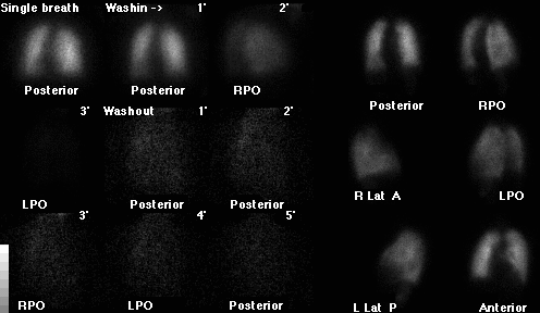
Ventilation-perfusion scintigraphy, 1/98, with ventilation
images at the left, and perfusion images at the right. Carefully examine the RPO and
LPO perfusion images.
View main image(vq) in a separate image viewer
View second image(vq).
Ventilation-perfusion scintigraphy, 8/92
View third image(xr).
Portable chest, 1/26/98 (top) and PA chest, 8/10/92 (bottom), filmed to
emphasize RUL findings
Full history/Diagnosis is available below
Diagnosis: Right-to-left Shunt Secondary to RUL Pulmonary Arteriovenous Malformation
Full history:
68-year-old woman who presents with progressive dyspnea, congestive
heart failure, and new onset atrial fibrillation. The patient's
peripheral arterial oxygenation was reported to be in the 40 mmHG
range.
Radiopharmaceutical:
13.4 mCi Xe-133 gas by inhalation and 4.3 mCi Tc-99m MAA intravenously
Findings:
The ventilation-perfusion pulmonary scintigraphy dated 1/26/98
demonstrates a subtle small ventilation defect in the right apex, with a
matched small perfusion defect in the right apex (best seen on
the RPO and right lateral images). These findings are more apparent on
the prior ventilation-perfusion scintigraphy of 8/10/92, and correspond
to an ill-defined opacity in the right upper lobe on the comparison
portable chest radiograph of 1/26/98. A similar opacity was noted on
the chest radiograph of 8/10/92. Both the ventilation-perfusion scintigraphies
were interpreted as low likelihood ratio for pulmonary embolism.
In addition to the small matched right apical ventilation-perfusion
defect, the perfusion studies also demonstrate
activity in the kidneys. No activity is identified in the thyroid,
salivary glands, or in the stomach. These findings are most indicative
of a right-to-left shunt.
Discussion:
While the emphasis on ventilation-perfusion scintigraphy is to evaluate
for ventilatory-perfusion mismatches, it is equally important to
be aware of extrapulmonary activity. The presence of extrapulmonary
activity on perfusion scintigraphy indicates the presence
of free Tc-99m pertechnetate or an intra- or extracardiac right-to-left
shunt. In a right-to-left shunt, radiopharmaceutical
will accumulate in systemic capillary beds, such as the kidneys, brain,
and spleen. In contrast, free Tc-99m pertechnate will accumulate most
prominently in the stomach, thyroid, salivary glands, and kidneys. An
image of the head is often helpful to distinguish between a right-to-left
shunt and free Tc-99m pertechnetate, as intracerebral activity is
present with a right-to-left shunt whereas localization of activity to
the scalp and cerebral sinuses is suggestive of free Tc-99m pertechnetate.
The diagnosis of a right-to-left shunt is important, as this
may be the explanantion for the patient's respiratory symptoms
that prompted the initial investigation for pulmonary thromboembolic
disease. Further investigation to determine whether a right-to-left
shunt is intracardiac or extracardiac (such as due to a pulmonary
arteriovenous malformation or right-to-left shunting from severe
endstage liver disease), usually will include a transthoracic echocardiogram
with bubble study as was done in this case. The findings on the
ventilation-perfusion scintigraphy in conjunction with the chronic
ill-defined right upper lobe opacity and the results of the
bubble study strongly suggest the presence of an intrapulmonary
right-to-left shunt, which was confirmed on pulmonary arteriography.
Followup:
The patient subsequently underwent a transthoracic echocardiogram which
demonstrated evidence of an extracardiac right-to-left shunt by bubble
study. A pulmonary arteriogram was then performed, which
revealed a right upper lobe pulmonary arteriovenous malformation
(see followup image below).
No other pulmonary arteriovenous malformations were
identified. Moderate to severe pulmonary arterial hypertension was
also noted, with pulmonary arterial pressure measurements of 63 mmHg
systolic, 22 mmHG diastolic, and 45 mmHG mean.
On the following day, the patient underwent successful coil embolization of the
right upper lobe arteriovenous malformation. Selective right
pulmonary arteriogram following coil embolization demonstrated no residual
flow within the arteriovenous malformation.
The right upper lobe arteriovenous malformation was felt to be
an incidental finding in this patient who does not have a history of
Osler-Weber-Rendu.
View followup image(an).
Selected images from selective right pulmonary arteriogram (left),
and following coil embolization of RUL arteriovenous malformation (right).
Major teaching point(s):
This case emphasizes the importance of evaluating for extrapulmonary
activity on perfusion scintigraphy.
A matched ventilation-perfusion defect with a corresponding radiographic
parenchymal opacity, as illustrated in this case, is nonspecific.
The most common differential diagnosis is pneumonia/infection. Less likely
considerations would include chronic mucous plugging and pulmonary
infarct. In this case, the chronicity of the right upper lobe opacity
in addition to the chronicity of the ventilation-perfusion findings would
make the likelihood of pulmonary embolism low.
Differential Diagnosis List
The presence of extrapulmonary
activity on perfusion scintigraphy typically indicates the presence of a
right-to-left shunt or free Tc-99m pertechnetate. Less commonly,
it may be due to recent administration of tracer from another nuclear
medicine study.
ACR Codes and Keywords:
References and General Discussion of Ventilation Perfusion Scintigraphy (Anatomic field:Lung, Mediastinum, and Pleura, Category:Normal, Technique, Congenital Anomaly)
Search for similar cases.
Edit this case
Add comments about this case
Return to the Teaching File home page.
Case number: vq026
Copyright by Wash U MO

