Case Author(s): Stephanie P.F. Yen, M.D. and Mark A. Mintun, M.D. , 10/24/97 . Rating: #D3, #Q4
Diagnosis: Massive bilateral pulmonary embolism
Brief history:
71-year-old female who presents with acute shortness of breath.
Images:
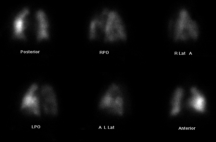
Perfusion scintigraphy.
View main image(vq) in a separate image viewer
View second image(vq).
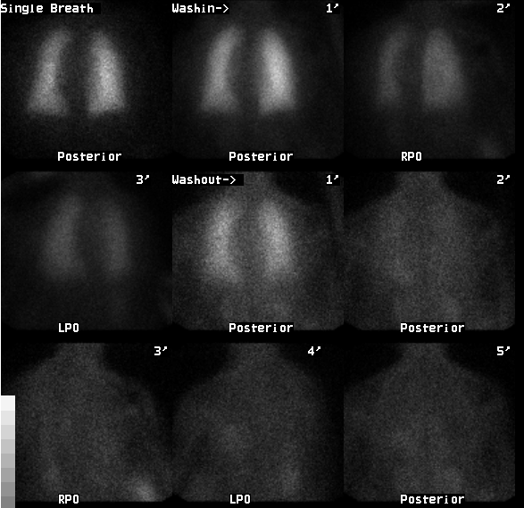
Ventilation scintigraphy.
View third image(xr).
Portable AP chest radiograph.
Full history/Diagnosis is available below
Diagnosis: Massive bilateral pulmonary embolism
Full history:
71-year-old female with history of pulmonary embolism and myasthenia
gravis who presents with acute shortenss of breath following a
near syncopal episode. Recent transesophageal echocardiogram performed
unexpectedly demonstrated intraluminal filling defect within the pulmonary artery,
suggestive of pulmonary embolism. A ventilation-perfusion pulmonary
scintigraphy study was requested for further evaluation.
Radiopharmaceutical:
16.4 mCi Xe-133 gas by inhalation and 4.4 mCi Tc-99m MAA, intravenously
Findings:
The ventilation study shows a uniform distribution of activity on the
single-breath and washin images. Minimal Xe-133 retention at the left
lung base is demonstrated during the washout phase. Perfusion
scintigraphy demonstrates
perfusion defects in the apicoposterior segment of the left upper lobe
as well as in the
posterobasal segment of the right lower lobe. These were noted on a
prior ventilation-perfusion study of 6/23/92 (not shown)
and are unchanged. However, the current study also demonstrates lobar
areas of relative hypoperfusion involving the entire left upper lobe,
left
lower lobe, and right lung. The lingula appears relatively
hyperperfused. The portable chest radiograph demonstrates no pulmonary
infiltrates or large pleural effusions. The scintigraphic findings are
worrisome for massive partially occluding bilateral pulmonary emboli.
Discussion:
In the evaluation of acute pulmonary embolism, regional areas of
decreased
ventilation leading to hypoxia-induced regional pulmonary
vasoconstriction typically occurs, resulting in regional hypoperfusion.
This is thought to occur due to the release of serotonin, prostaglandins,
and angiotensin II in response to hypoxia. In contrast, the underlying
etiology and physiology responsible for the occasional appearance of focal zones of
relative hyperperfusion ("hot spots") on perfusion scintigraphy is not
clearly understood. The zones of apparent hyperperfusion may reflect
true local vasodilatation or preserved perfusion in an area of normal
lung adjacent to abnormal lung regions.
To investigate the frequency and significance of hot spots on perfusion
scintigraphy, Meigan et al. (1989) reviewed 72 patients who underwent
both ventilation-perfusion scintigraphy and pulmonary
angiography. Hot spots were seen in 18% of the perfusion studies, the
majority of which involved less than one pulmonary segment. Most of the
focal zones of hyperperfusion corresponded to areas of atelectasis on
chest radiograph. It is postulated that the failure of hypoxic
vasoconstriction in poorly ventilated lung results in a relative
increase of perfusion in the atelectatic lung. In 3 patients with massive
bilateral pulmonary emboli demonstrated
scintigraphically and documented by angiography, as illustrated in this
case, the focal zones of hyperperfusion represented areas of preserved
normal perfusion.
Reference: Meignan M, Palmer EL, Waltman AC, Strauss HW. Zones of
increased perfusion (hot spots) on perfusion lung scans: Correlation
with pulmonary arteriograms. Radiology 1989; 173:47-52.
Followup:
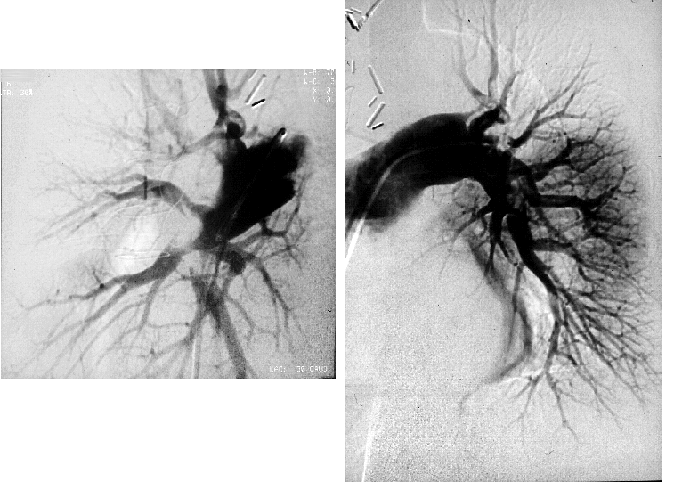
The patient subsequently underwent a pulmonary arteriogram later the same
day, 10/17/97, which revealed bilateral large lower lobe and smaller
upper lobe pulmonary emboli.
View followup image(an).
Selective images from pulmonary arteriogram: right pulmonary
arterial injection (right) and left pulmonary arterial injection
(left).
Major teaching point(s):
Focal zones of hyperperfusion or "hot spots" on perfusion
scintigraphy may represent areas of atelectasis. More importantly,
hot spots may signify a patient with a large embolic burden, with
the hot spots representing regions of relatively normal
perfusion adjacent to hypoperfused, embolized lung.
ACR Codes and Keywords:
References and General Discussion of Ventilation Perfusion Scintigraphy (Anatomic field:Lung, Mediastinum, and Pleura, Category:Other generalized systemic disorder)
Search for similar cases.
Edit this case
Add comments about this case
Return to the Teaching File home page.
Case number: vq025
Copyright by Wash U MO