Case Author(s): Michael Quinn, M.D. and Keith Fischer, M.D. , 09/05/97 . Rating: #D2, #Q4
Diagnosis: Physiologic shunt (Perfused but non-ventilated lung.)
Brief history:
18 yo. male with SOB and hypoxia
Images:

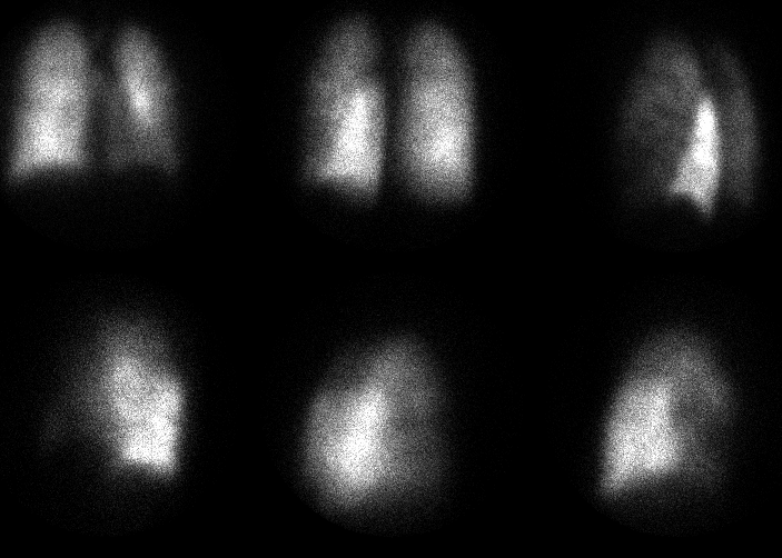
Tc-99mDTPA aerosol ventilation images
(Antrior, posterior, lpo, rpo)
View main image(vq) in a separate image viewer
View second image(vq).
Tc99mMAA perfusion images
(Ant, Post, LPO, LLat, RPO, RLat)
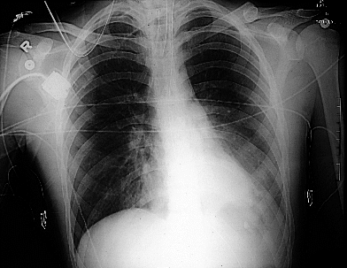
View third image(xr).
Chest radiograph
Full history/Diagnosis is available below
Diagnosis: Physiologic shunt (Perfused but non-ventilated lung.)
Full history:
18 year old male involved in MVA 2 days prior to this study. During
his hospital admission he developed shortness of breath and was found to
be hypoxic. The referring service was concerned about possible
pulmonary embolism.
Radiopharmaceutical:
Tc99mDTPA aerosol for ventilation, Tc99mMAA for perfusion
Findings:
On ventilation images, there is decreased activity at the left lung
base. The chest radiograph is remarkable for an area of retrocardiac
consolidation corresponding to the region of decreased ventilation.
The perfusion images demonstrate relative increased perfusion to the
entire left lower lobe.
Discussion:
This study is an excellent example of a physiologic shunt causing
clinical symptoms. Such shunts result when non-ventilated regions of
lung continue to remain perfused secondary to failure of the usual
reflex vasoconstriction associated with hypoventilation.
In this patient, the decreased left lung base ventilation was thought
to be secondary to an obstructing mucous plug. The lung distal to this
obstruction continued to receive blood flow, causing non-oxygenated
blood to enter the systemic circulation and resulting in hypoxia. There
was no evidence for pulmonary embolism. When this patient was rolled
onto his right side, this decreased the size of the shunt and the
patients hypoxia immediately and drastically improved. Treatment
consisted of relieving the bronchial obstruction.
Differential Diagnosis List
Mucous plug, endobronchial lesion, extrinsic bronchial compression.
ACR Codes and Keywords:
References and General Discussion of Ventilation Perfusion Scintigraphy (Anatomic field:Lung, Mediastinum, and Pleura, Category:Organ specific)
Search for similar cases.
Edit this case
Add comments about this case
Return to the Teaching File home page.
Case number: vq024
Copyright by Wash U MO