Case Author(s): Matt Jaksha, M.D. and Jerold Wallis, M.D. , 8/1/97 . Rating: #D2, #Q3
Diagnosis: Mucous plug
Brief history:
41 year old male in ICU with new onset of atrial fibrillation.
Rule out pulmonary embolus.
Images:
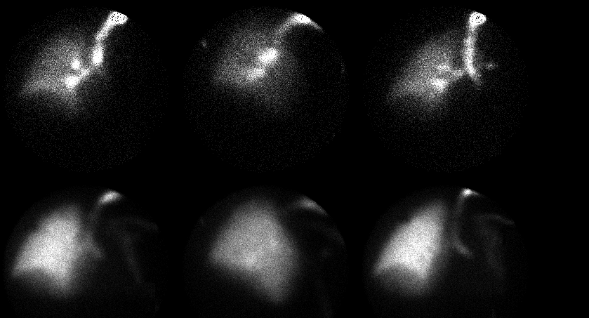
Anterior, RAO and LAO ventilation(top) and perfusion(bottom) images.
View main image(vq) in a separate image viewer
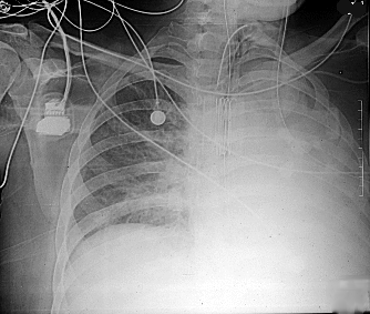
View second image(xr).
CXR from the same day.
Full history/Diagnosis is available below
Diagnosis: Mucous plug
Full history:
This 41 year old male was admitted with necrotizing pancreatitis.
Subsequent complications include abdominal abscess. He now presents
with new onset of atrial fibrillation. Ventilation-perfusion lung
scintigraphy is requested to rule out pulmonary emboli.
Radiopharmaceutical:
Tc-99m DTPA aerosol and Tc-99m MAA
Findings:
The ventilation and perfusion of the right lung are normal. There is
almost a complete lack of ventilation and perfusion to the left lung.
The comparison chest radiograph shows diffuse opacification of the left
hemithorax with apparent volume loss.
Discussion:
Although standard interpretation criteria places perfusion abnormalities
with corresponding radiographic abnormalities into the "intermediate"
category, pulmonary emboli would be unlikely to result in the
scintigraphic pattern seen in this case. Mucous plugging of the left
mainstem bronchus with reflex vasoconstriction and volume
loss is a more likely explanation for this appearance.
Pneumonia and/or pleural effusion are possible, though these
alone would not
explain the left-sided volume loss.
Followup:
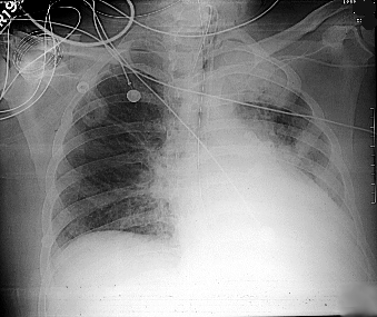
After the patient was suctioned via the endotracheal tube, his condition
improved significantly. The next day's chest radiograph showed
improvement in aeration of the left lung.
View followup image(xr).
CXR from the following day with improved aeration on the left.
Major teaching point(s):
Lack of ventilation to an entire lung is unlikely to result from a
pulmonary embolus. Mucous plugging in an intubated patient can cause
this finding, and appropriate clinical measures (e.g. suctioning
or bronchoscopy) should be taken.
Differential Diagnosis List
Foreign body,
mediastinal/hilar mass,
any lesion obstructing the airway.
ACR Codes and Keywords:
References and General Discussion of Ventilation Perfusion Scintigraphy (Anatomic field:Lung, Mediastinum, and Pleura, Category:Organ specific)
Search for similar cases.
Edit this case
Add comments about this case
Return to the Teaching File home page.
Case number: vq023
Copyright by Wash U MO