Case Author(s): Scott Winner, M.D. and Henry Royal, M.D. , 12/06/96 . Rating: #D3, #Q4
Diagnosis: Intrapulmonary right to left shunt
Brief history:
75-year old man with
increasing shortness of breath and dyspnea.
Images:

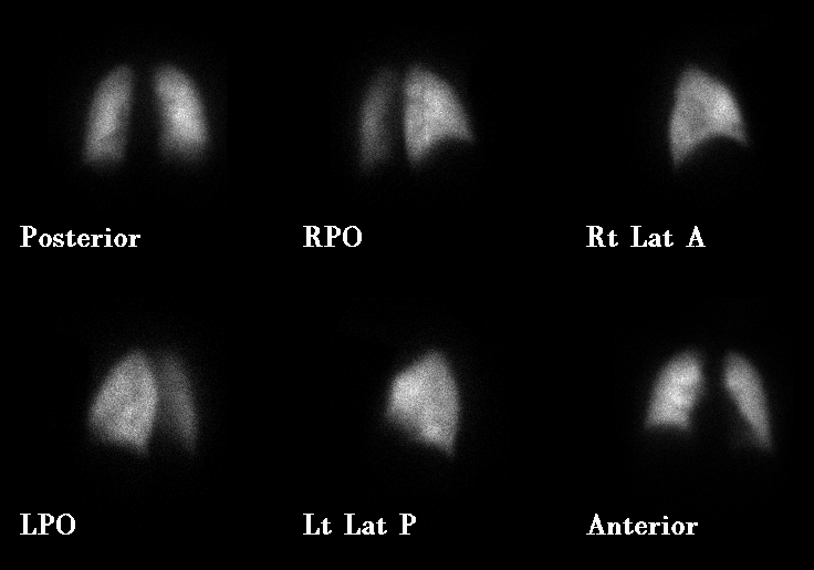
Pulmonary Perfusion Images
View main image(vq) in a separate image viewer
View second image(vq).
Pulmonary Ventilation Images
View third image(xr).
Chest radiograph
Full history/Diagnosis is available below
Diagnosis: Intrapulmonary right to left shunt
Full history:
75-year old man with
chronic renal insufficiency and congestive heart
failure with increasing shortness of breath and
dyspnea. A comparison chest radiograph dated
12-2-96 demonstrates an infiltrate in the left
mid lung consistent with pneumonia.
Radiopharmaceutical:
12.9 mCi Xe-133
gas by inhalation and 4.2 mCi Tc-99m MAA
i.v.
Findings:
The Xe-133 ventilation images
show markedly reduced ventilation to the left
lung. The perfusion images show only
minimally reduced perfusion to the left lung.
Quantitative analysis demonstrates that the
right lung receives 59% and the left lung
receives 41% of total pulmonary blood flow.
The right lung contributes 84% and the left
lung contributes 16% of total pulmonary
ventilation. The above subjective and
quantitative findings are consistent with a
significant intrapulmonary right-to-left
physiologic shunt involving the left lung.
Discussion:
In patients with pneumonia, lung
scintigraphy commonly demonstrates matched
ventilation and perfusion abnormalities.
Regional alveolar hypoxia in areas of infected
lung usually results in reflex hypoxic
pulmonary vasoconstriction. This effectively
shunts blood to normal well ventilated areas of
lung. This physiology results in matched
ventilation/perfusion defects.
Less commonly in patients with pneumonia,
a reverse mismatch is present (perfusion is
maintained and ventilation is reduced or
absent). This is thought to be related to local
release of inflammatory mediators with
vasodilatory properties which overcome the
hypoxic reflex vasoconstriction. The result is
an intrapulmonary right to left physiologic
shunt which can render the patient hypoxic and
short of breath. Pulmonary emboli do not
present with reverse mismatches.
References:
Li et al. Scintigraphic
Appearance in Patients With Pulmonary
Infection and Lung Scintigrams of Intermediate
or Low Probability for Pulmonary Embolism.
Clin Nucl Med 19:1091-1093, 1994.
Followup:
None.
Major teaching point(s):
None.
Differential Diagnosis List
Reverse
mismatch can be caused by pneumonia,
bronchial obstruction, collapse, pleural
effusion, metabolic alkalosis and pulmonary
hypertension.
ACR Codes and Keywords:
- General ACR code: 62
- Lung, Mediastinum, and Pleura:
6.217 "Opportunistic infection"
References and General Discussion of Ventilation Perfusion Scintigraphy (Anatomic field:Lung, Mediastinum, and Pleura, Category:Inflammation,Infection)
Search for similar cases.
Edit this case
Add comments about this case
Return to the Teaching File home page.
Case number: vq022
Copyright by Wash U MO

