Case Author(s): Anton J. Johnson, M.D., Ph.D. and Henry D. Royal, M.D. , 11/8/96 . Rating: #D2, #Q4
Diagnosis: Pulmonary Embolism
Brief history:
43 year old woman with a right lung transplant and worsening dyspnea.
Images:

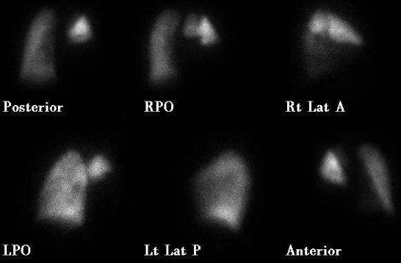
Perfusion images (10/30/96).
View main image(vq) in a separate image viewer
View second image(vq).
Ventilation images (10/30/96).
View third image(xr).
Chest film (10/30/96).
View fourth image(an).
Pulmonary angiogram (10/31/96).
Full history/Diagnosis is available below
Diagnosis: Pulmonary Embolism
Full history:
43 year old woman who, three years earlier, underwent right lung
transplantation for primary pulmonary hypertension. She now presents
with worsening dyspnea following a syncopal episode.
Radiopharmaceutical:
4.2 mCi Tc-99m MAA i.v. and 10.4 mCi Xe-133 gas by inhalation.
Findings:
The initial perfusion images (10-30-96) demonstrate essentially no
tracer activity in the right lower and right middle lobes as well as
decreased perfusion to the apical and posterior segments of the right
upper lobe. The ventilation portion of the study reveals a defect in
the right lung base and there is opacification in the right base on the
comparison chest radiograph (10-30-96).
Discussion:
The right lung perfusion defect is substantially larger than the
corresponding ventilatory defect and radiographic opacity. In addition,
there are no right upper lobe ventilation abnormalities to match the
perfusion defects in the apical and posterior segments of the right
upper lobe. Regardless of which interpretive scheme you use, these
findings are consistent with a high likelihood ratio for pulmonary embolism.
A pulmonary angiogram was performed in order to determine whether or not
the lesions would be amenable to embolectomy. The angiogram (10/31/96)
shows a large round filling defect in the right main pulmonary artery
with some contrast getting by to partially opacify vessels supplying the
right upper lobe.
Followup:
The patient underwent surgical embolectomy of a large thrombus in her
right main pulmonary artery. The thrombus completely filled the arterial
lumen except for a tiny channel that allowed some blood flow to the
right upper lobe. A caliber change at the arterial anastomosis was felt
to be the site of origin of the thrombus.
A follow-up (post-embolectomy) portable perfusion/aerosol study reveals
marked improvement in the perfusion to the right lower lobe.
View followup image(vq).
Follow-up portable perfusion/aerosol study (11/1/96).
ACR Codes and Keywords:
References and General Discussion of Ventilation Perfusion Scintigraphy (Anatomic field:Lung, Mediastinum, and Pleura, Category:Organ specific)
Search for similar cases.
Edit this case
Add comments about this case
Return to the Teaching File home page.
Case number: vq020
Copyright by Wash U MO

