Case Author(s): Scott Winner, M.D. and Jerold Wallis, M.D. , 9/27/96 . Rating: #D2, #Q3
Diagnosis: Pulmonary Embolism
Brief history:
41-year old woman with
shortness of breath.
Images:

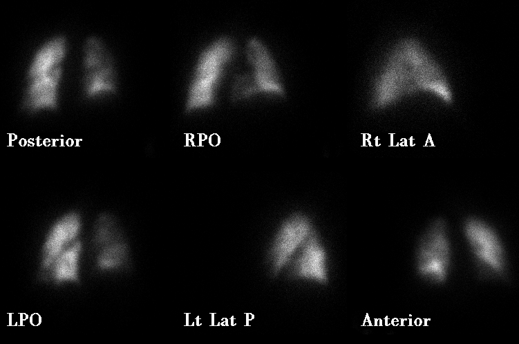
Perfusion images of the lungs
View main image(vq) in a separate image viewer
View second image(vq).
Ventilation images of the lungs
View third image(ct).
CT of the chest
The chest radiograph (not shown) was without infiltrates or effusions.
Full history/Diagnosis is available below
Diagnosis: Pulmonary Embolism
Full history:
41-year old woman with
history of metastatic breast cancer, increased
shortness of breath, and decreased 02
saturation.
Radiopharmaceutical:
4.3 mCi Tc-99m
MAA iv and 19.7 mCi Xe-133 gas by inhalation
Findings:
Decreased perfusion is seen
to the right lung (particularly evident in the right lower
lobe on the RPO image) and (after allowing for the heart) a moderate segmental
defect in the anterior portion of the left lower lobe (best
seen on the left lateral image). In the absence of significant
ventilation or radiographic abnormalities, this yields a
high likelihood ratio for pulmonary embolism.
CT findings are discussed in the "followup" section below.
Discussion:
Isolated lobar or whole lung decreased perfusion is relatively
atypical in pulmonary embolism, and should raise the suspicion of
other central obstructing lesions (e.g. hilar tumor mass, fibrosing
mediastinitis). In this case, an additional defect is present in the
contralateral lung to increase the suspicion for pulmonary embolism.
Computed tomography may be the confirmatory test of choice in this
setting, since it can both evaluate for hilar mass and detect
central obstructing clot.
References: Remy-Jardin M, et al.,
Diagnosis of Pulmonary Embolus with Spiral CT:
Comparison with Pulmonary Angiography and
Scintigraphy. Radiology 1996; 200:699-706.
Followup:
CT scan of the chest dated 8-
31-96 showed a large central pulmonary
embolus in the right interlobar pulmonary
artery with additional filling defects seen in the
left lower lobe pulmonary artery consistent
with pulmonary embolism.
Major teaching point(s):
When a perfusion
abnormality involves an entire lung or lobe,
one should consider other etiologies for the
perfusion defect. This is a somewhat unusual
presentation for pulmonary emboli.
Differential Diagnosis List
Central tumor
obstructing the pulmonary artery, mediastinal
fibrosis, vascular anomaly (pediatric patients)
and large central pulmonary embolus.
ACR Codes and Keywords:
- General ACR code: 67
- Lung, Mediastinum, and Pleura:
6.721 "Pulmonary embolization without infarction"
References and General Discussion of Ventilation Perfusion Scintigraphy (Anatomic field:Lung, Mediastinum, and Pleura, Category:Organ specific)
Search for similar cases.
Edit this case
Add comments about this case
Read comments about this case
Return to the Teaching File home page.
Case number: vq019
Copyright by Wash U MO

