Case Author(s): Samuel Wang, M.D. and Barry A. Siegel, M.D. , . Rating: #D3, #Q3
Diagnosis: Pulmonary sling.
Brief history:
18-year-old male patient with
sickle-cell anemia who presented with chest pain.
Images:
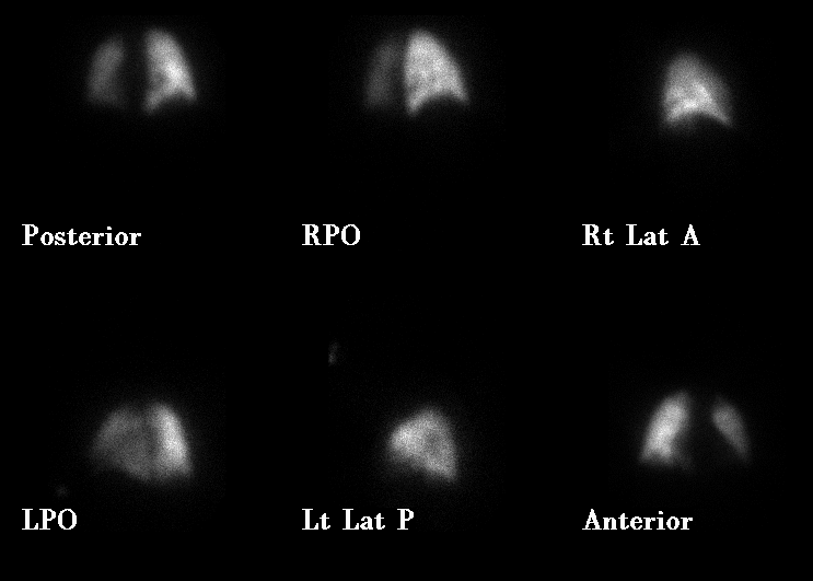
Perfusion images.
View main image(vq) in a separate image viewer
View second image(vq).
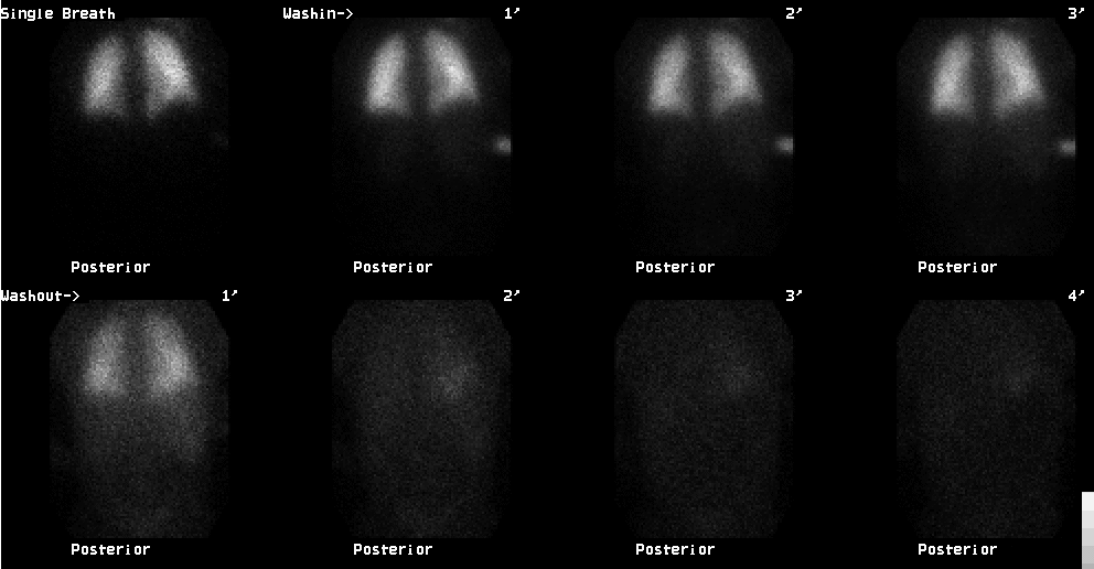
Posterior ventilation images.
View third image(xr).
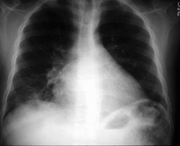
PA chest radiograph.
View fourth image(xr).
Lateral chest radiograph.
Full history/Diagnosis is available below
Diagnosis: Pulmonary sling.
Full history:
18-year-old male patient with
sickle cell anemia who presented with atypical mid
sternal chest pain after being seated for multiple
hours. He was noted to be hypoxic on room air.
Radiopharmaceutical:
Xe-133 gas and Tc-99m
MAA
Findings:
The ventilation images demonstrate
Xe-133 retention at the right lung base. The perfusion
images demonstrate decreased perfusion to the right
lung base, matching the ventilatory abnormality.
Additionally, there is diffuse hypoperfusion of the
entire left lung. The chest radiograph demonstrates
mild cardiomegaly with H-shaped vertebrae and bony
sclerosis consistent with the patient's sickle cell
anemia. No confluent infiltrates or pleural effusions
were identified.
Discussion:
The matched perfusion defects in
the right lower lung were felt to be most likely due to
focal obstructive disease. The differential diagnosis
offered for unilateral decreased perfusion without
corresponding ventilatory abnormality included an
unusual presentation of pulmonary thromboembolism (acute or
chronic in situ thrombosis related to the patient's
sickle cell anemia), extrinsic compression of the left
pulmonary artery due to tumor or inflammatory lymphadenopathy, pulmonary artery
hypoplasia, or vasculitis such as Takayasu's arteritis.
Followup:
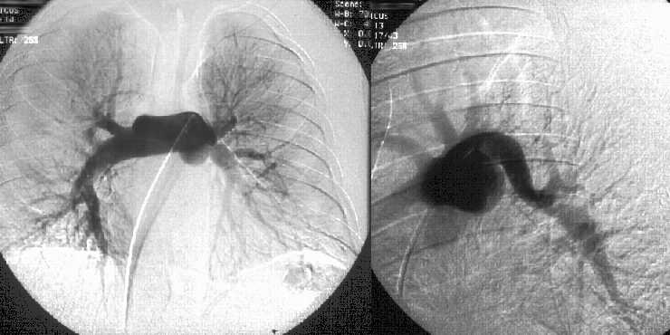
A pulmonary arteriogram was
performed and demonstrated a pulmonary vascular
sling with the aberrant left pulmonary artery arising
from the right pulmonary artery. An area of moderate
compression was seen in the left pulmonary artery just
proximal to the bifurcation of the upper and lower lobe
vessels. The presence of a pulmonary sling was also
confirmed by a CT study. Review of the patient's chest
radiograph demonstrates the posterior tracheal
indentation and anterior esophageal impression seen
with an aberrant left pulmonary artery.
A pulmonary sling, also known as aberrant left
pulmonary artery, represents failure of development of
the left 6th aortic arch followed by development of a
collateral branch of the right pulmonary artery to
supply the left lung. Typically, the left pulmonary
artery passes above the right mainstem bronchus and
between the trachea and esophagus to supply the left
lung. This anomaly can cause obstructive pulmonary
disease with stridor being the most common
presentation.
View followup image(an).
Pulmonary arteriogram
ACR Codes and Keywords:
References and General Discussion of Ventilation Perfusion Scintigraphy (Anatomic field:Heart and Great Vessels, Category:Normal, Technique, Congenital Anomaly)
Search for similar cases.
Edit this case
Add comments about this case
Return to the Teaching File home page.
Case number: vq018
Copyright by Wash U MO