Case Author(s): Charles Pirngle, M.D., Tom R. Miller, M.D., Ph.D. , 02/13/96 . Rating: #D2, #Q3
Diagnosis: Fibrosing Mediastinitis
Brief history:
Shortness of breath
Images:
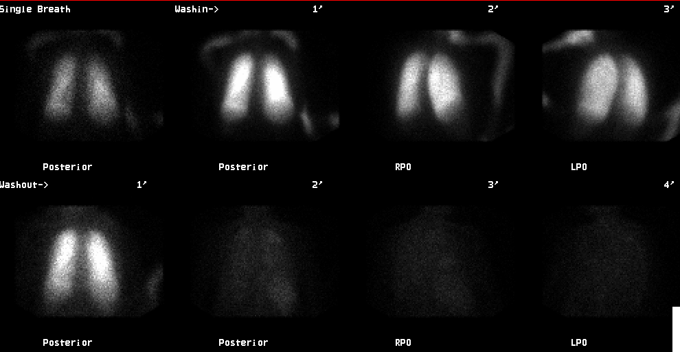
Posterior ventilation images
View main image(vq) in a separate image viewer
View second image(vq).
Lung perfusion images
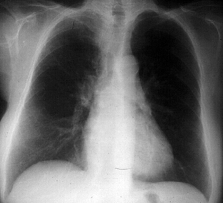
View third image(xr).
PA chest radiograph
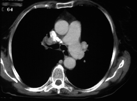
View fourth image(ct).
Transverse CT image through the mediastinum
Full history/Diagnosis is available below
Diagnosis: Fibrosing Mediastinitis
Full history:
71-year old woman with a right
upper lobe infiltrate and a past history of pulmonary
embolism. She now has shortness of breath.
Radiopharmaceutical:
20.5 mCi Xe-133 gas by
inhalation and 4.1 mCi Tc-99m MAA i.v.
Findings:
The Xe-133 ventilation images show
mildly reduced ventilation in the right upper lobe
manifested by decreased single-breath and washin
activity with mild retention during washout. The
perfusion images show a matching defect involving
the entire right upper lobe. Also, according to the
report of an outside study from one year earlier, there
has been no change in the appearance of the
ventilation-perfusion study. The chest radiograph
demonstrates narrowing of the distal trachea and also
possibly the right mainstem bronchus with streaky
opacities in the right upper lobe, suggesting an area of
infiltrate or atelectasis. Also, a recent CT scan of the
chest demonstrated right hilar adenopathy with
coarse calcification and volume loss in the right upper
lobe.
Discussion:
Fibrosing mediastinitis is the
most likely etiology for the computed tomographic,
radiographic, and ventilation-perfusion abnormalities.
The most frequent cause of fibrosing mediastinitis is
granulomatous disease, with
histoplasmosis the likely offender in the Midwest.
Tuberculosis and actinomycosis may also result in the
same findings. Other causes of fibrosing mediastinitis
include autoimmune disease and methysergide. The
characteristic scintigraphic findings, present in this
case, are decreased to absent perfusion involving a
large area of lung with normal or near-normal
ventilation.
Major teaching point(s):
It would be unusual for
pulmonary embolism to present as a single lobar or whole lung
defect. Because PE occur multiply, the single defect suggests other diagnoses.
Differential Diagnosis List
In addition to fibrosing mediastinitis, the possibilities include a central obstructing mass, such as bronchogenic
carcinoma, Swire-James syndrome, and, unlikely,
embolus to the upper lobe pulmonary artery.
ACR Codes and Keywords:
References and General Discussion of Ventilation Perfusion Scintigraphy (Anatomic field:Lung, Mediastinum, and Pleura, Category:Inflammation,Infection)
Search for similar cases.
Edit this case
Add comments about this case
Read comments about this case
Return to the Teaching File home page.
Case number: vq017
Copyright by Wash U MO