Case Author(s): J. Philip Moyers, M.D. and Henry D. Royal, M.D. , 12/5/95 . Rating: #D2, #Q3
Diagnosis: Idiopathic bronchial stenosis
Brief history:
Withheld
Images:
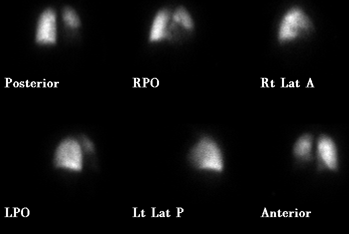
Perfusion images from VQ scan
View main image(vq) in a separate image viewer
View second image(vq).
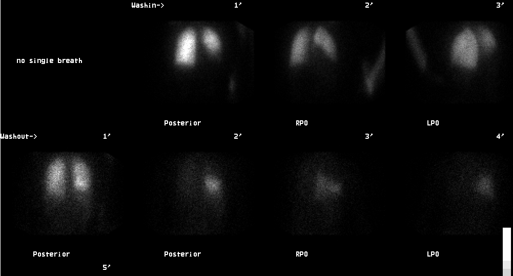
Ventilation images from VQ scan
View third image(xr).
Pa Chest Radiograph
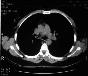
View fourth image(ct).
Axial section from Chest CT, mediastinal windows
Full history/Diagnosis is available below
Diagnosis: Idiopathic bronchial stenosis
Full history:
This patient has known right
bronchus intermedius bronchial stenosis and a history
of tracheal stenosis in 1988. Recent bronchoscopy
demonstrated the stenosis in the bronchus
intermedius and this stenosis was dilated at the time
of the bronchoscopy.
Radiopharmaceutical:
11.3 mCi Xe-133 gas by
inhalation and 3.6 mCi Tc-99m MAA i.v.
Findings:
Poor washin of the Xe-133 is
demonstrated on the washin images in the right
middle and lower lobes. This corresponds well with
the decreased perfusion demonstrated in these regions
on the perfusion images. Delayed washout and
prolonged retention of Xe-133 is demonstrated during
the washout phase of the ventilation portion of the
examination.
Discussion:
This patient carries a diagnosis of
idiopathic bronchial stenosis. The patient is status
post at least two dilatation attempts of the bronchus
intermedius. CT examination (included)
demonstrates narrowing of the bronchus intermedius
without evidence for extraluminal mass. There is no
evidence for mass involving the right-sided intralobar
artery. This case demonstrates the physiologic
hypoxic vasoconstriction that can occur in areas of
lung that are not well ventilated. Decreased perfusion
is demonstrated on the perfusion images and on chest
radiograph (not included) relative oligemia is
demonstrated in the right lower lung zone. Lung
windows of the chest CT also demonstrate relative
oligemia of the right lower lung zone compared to the
left lower lung zone.
ACR Codes and Keywords:
References and General Discussion of Ventilation Perfusion Scintigraphy (Anatomic field:Lung, Mediastinum, and Pleura, Category:Organ specific)
Search for similar cases.
Edit this case
Add comments about this case
Read comments about this case
Return to the Teaching File home page.
Case number: vq015
Copyright by Wash U MO