Case Author(s): J. Philip Moyers, M.D. and Keith C. Fischer, M.D. , 12/5/95 . Rating: #D1, #Q3
Diagnosis: High likelihood ratio for pulmonary
embolism
Brief history:
Shortness of breath
Images:

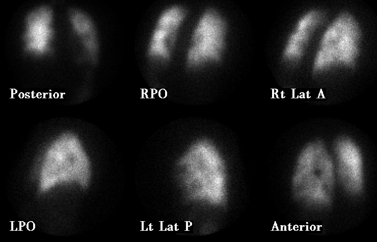
Initial perfusion images
View main image(vq) in a separate image viewer
View second image(vq).
Initial ventilation images (portable)
View third image(pe).
Perfusion images obtained the following day
View fourth image(xr).
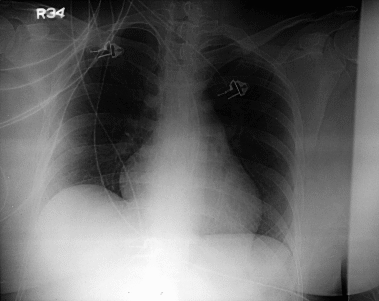
portable supine AP chest radiograph
Full history/Diagnosis is available below
Diagnosis: High likelihood ratio for pulmonary
embolism
Full history:
36-year old woman with calf
pain, shortness of breath, and chest pain which began
two days ago.
Radiopharmaceutical:
Less than 2 mCi Tc-
99m DTPA aerosol by inhalation and 5.3 mCi Tc-99m
MAA i.v.
Findings:
Perfusion images obtained on 11-9-95
demonstrate multiple segmental perfusion defects
throughout both lungs. Perfusion images obtained
two days later demonstrate near total resolution of
these multiple segmental perfusion defects. Interval
treatment included intravenous urokinase performed
secondary to marked cardiovascular compromise of
the patient due to these multiple pulmonary emboli.
Discussion:
Pulmonary embolism (PE) is diagnosed
in 1% of all hospitalized patients and seen in 15-60% of
autopsies. There are 600,000 new cases per year. The classic
clinical triad, which is seen in less than one-third of patients,
includes hemoptysis, pleural friction rub, and
thrombophlebitis. While the major cause of PE is deep venous
thrombosis (DVT), only 10-30% of patients with fatal PE have
symptomatic DVT. Chest radiographs are notoriously
insensitive and nonspecific in clinically suspected pulmonary
embolism. Ventilation-perfusion scintigraphy is the diagnostic
test of choice.
ACR Codes and Keywords:
References and General Discussion of Ventilation Perfusion Scintigraphy (Anatomic field:Lung, Mediastinum, and Pleura, Category:Organ specific)
Search for similar cases.
Edit this case
Add comments about this case
Read comments about this case
Return to the Teaching File home page.
Case number: vq014
Copyright by Wash U MO