Case Author(s): Michael C. Roarke, M.D., Barry A. Siegel, M.D. , 11/24/95 . Rating: #D3, #Q4
Diagnosis: Pulmonary Embolism
Brief history:
66-year-old man with
stage IV esophageal carcinoma admitted with right
pleuritic flank pain. Arterial blood gas analysis
revealed a pH of 7.40, p02 of 54 mm Hg, and pC02 of
36 mm Hg.
Images:

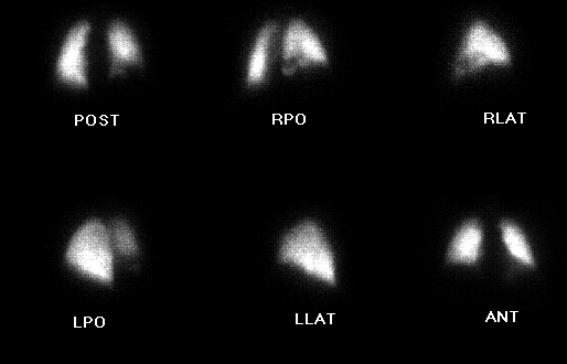
Xe-133 ventilation images.
View main image(vq) in a separate image viewer
View second image(vq).
Perfusion images.
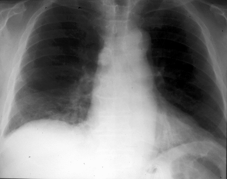
View third image(xr).
PA chest radiograph.
View fourth image(an).
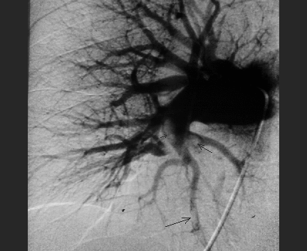
Pulmonary arteriogram.
Full history/Diagnosis is available below
Diagnosis: Pulmonary Embolism
Full history:
66-year old gentleman with
stage IV esophageal carcinoma admitted with right
pleuritic flank pain. Arterial blood gas analysis
revealed a pH of 7.40, p02 of 54 mm Hg, and pC02 of
36 mm Hg.
Radiopharmaceutical:
12.9 mCi Xe-133 gas by
inhalation and 4.4 mCi Tc-99m MAA i.v.
Findings:
The Xe-133 ventilation images
showed a washin defect involving the posterior and
lateral basal segments of the right lower lobe. No
abnormal Xe-133 retention was identified during the
washout phase. The perfusion images demonstrated
matching perfusion defects in the posterior and lateral
basal segments of the right lower lobe, which were
slightly more severe than the ventilatory
abnormalities. The chest radiographs revealed hazy
opacity in the right lower lobe, which matched the
ventilatory and perfusion abnormalities. This
examination was interpreted as representing an
intermediate likelihood ratio for pulmonary embolism.
The pulmonary arteriogram revealed multiple filling
defects within right lower lobe pulmonary arterial
branches consistent with pulmonary emboli.
Discussion:
In this case, the ventilation and
perfusion abnormalities are similar in location and
severity, although the perfusion defect is arguably
slightly more severe than the ventilatory defects.
The chest radiographs reveal an opacity in the
right lower lobe, which matches the ventilation and
perfusion abnormalities. This study would be
interpreted as indicative of an intermediate likelihood ratio or
intermediate probability for pulmonary embolism with both the PIOPED and
modified Biello criteria, given two perfusion defects
matching the ventilatory and chest radiographic
abnormalities. However, given the slightly more
severe perfusion defect compared with the ventilatory
abnormality, one might consider informing the
patient's physicians that the suspicion of pulmonary
embolism is slightly greater than if the defects were
all perfectly matched.
References: Palmer, Scott, Strauss.
Practical Nuclear Medicine, pg 202-203, 1992
Followup:
A follow-up ventilation-perfusion
study was performed three
months later, which revealed nearly complete resolution of the
perfusion defects seen on this examination.
ACR Codes and Keywords:
References and General Discussion of Ventilation Perfusion Scintigraphy (Anatomic field:Lung, Mediastinum, and Pleura, Category:Misc)
Search for similar cases.
Edit this case
Add comments about this case
Read comments about this case
Return to the Teaching File home page.
Case number: vq013
Copyright by Wash U MO