Case Author(s): J. Philip Moyers, MD and Farrokh Dehdashti, MD , 11/16/95 . Rating: #D4, #Q4
Diagnosis: Takayasu's Arteritis
Brief history:
This patient presents with left-
sided shoulder pain.
Images:
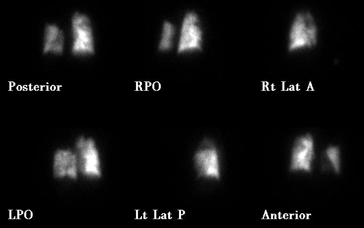
Projection images from a lung perfusion scan
View main image(pe) in a separate image viewer
View second image(vq).
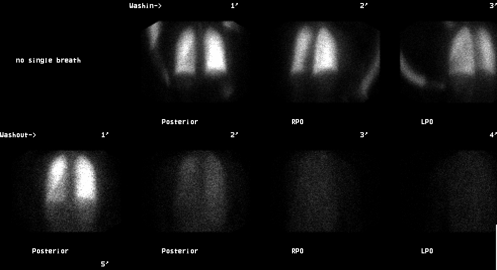
Projectional images from Xenon-133 ventilation portion of VQ scan
View third image(an).
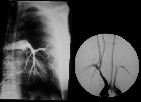
Anterior projection of right PA injection, LAO projection of arch aortogram
Full history/Diagnosis is available below
Diagnosis: Takayasu's Arteritis
Full history:
37-year old Oriental woman with left
shoulder pain and shortness of breath and known history of Takayasu's arteritis
Radiopharmaceutical:
17.1 mCi Xe-133 gas by
inhalation and 4.4 mCi Tc-99m MAA i.v.
Findings:
Overall decreased perfusion to the entire
left lung, especially the apical-posterior segment of the let
upper lobe. There is also a small perfusion defect in the
anterior segment of the right upper lobe (on the RPO view).
Discussion:
Takayasuąs arteritis, also known as
pulseless disease, is a chronic inflammatory panarteritis of
unknown etiology affecting predominantly large central
arteries, including the aorta (and its branches) and the
pulmonary arteries. 2.6 new cases per million are diagnosed
per year. This disease typically afflicts young Oriental
females (age range 15-41 years). The clinical signs and
symptoms may be divided in a pre-pulseless phase and a
pulseless phase. The pre-pulseless phase, which may last from
several months to a year, is characterized by weight loss,
fevers, myalgias, arthralgias, and other nonspecific symptoms.
The pulseless phase includes claudication, pulse deficits, bruits
(plus other signs and symptoms of limb ischemia), and renal
vascular hypertension.
The angiographic findings include both long, diffuse, and
short segmental irregular stenotic areas of the major branches
of the aorta near their origins. The thoracic aorta is more
commonly affected than the abdominal aorta. The
angiographic findings typically describe skip areas and
abundant collaterals. Fusiform aortic aneurysm and ectasias
are seen in 10-15%.
Followup:
Follow-up pulmonary angiogram proved Takayasu's arteritis.
Differential Diagnosis List
Takayasu's arteritis may be mistaken for pulmonary embolism as both conditions exhibit abnormal perfusion and normal ventilation on ventilation-perfusion scintigraphy.
ACR Codes and Keywords:
References and General Discussion of Ventilation Perfusion Scintigraphy (Anatomic field:Lung, Mediastinum, and Pleura, Category:Other generalized systemic disorder)
Search for similar cases.
Edit this case
Add comments about this case
Read comments about this case
Return to the Teaching File home page.
Case number: vq012
Copyright by Wash U MO