Case Author(s): Gregg Schubach/F. Dehdashti , 9/8/95 . Rating: #D2, #Q4
Diagnosis: Swyer-James syndrome
Brief history:
7-year old boy with history of
previous pneumonia.
Images:

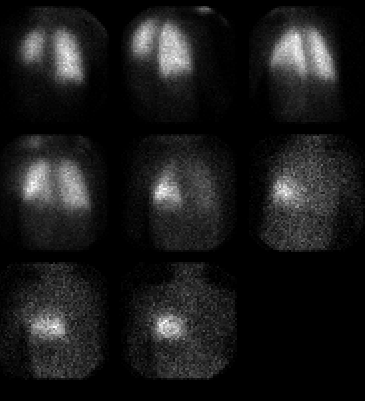
one minute sequential ventilation images
View main image(vq) in a separate image viewer
View second image(vq).
perfusion images
View third image(xr).
PA Chest Radiograph (contrast adjusted to accentuate lung markings)
Full history/Diagnosis is available below
Diagnosis: Swyer-James syndrome
Full history:
7-year old boy with history of
previous pneumonia. The patient1s recent chest
radiograph demonstrates diminished vascularity in
the left lower lung zone. The patient is currently
asymptomatic. The clinical and radiographic findings
are suggestive of Swyer-James syndrome.
Ventilation-perfusion scintigraphy was obtained to
assist in confirming this diagnosis.
Findings:
The Xe-133 washin ventilation
images demonstrate hypoventilation of the left lower
lung. The washout images show Xe-133 retention in
this region. This abnormality corresponds to the
region of radiographic abnormality. The perfusion
images show hypoperfusion in the left lower lung,
corresponding to both the ventilation and
radiographic abnormalities.
Discussion:
Swyer-James syndrome
(Macleod syndrome, unilateral hyperlucent lung) is
likely secondary to childhood adenoviral infection
with subsequent acute obliterative bronchiolitis
developing between 7-30 months post infection. At
the time of diagnosis, the patients are usually
asymptomatic but may experience dyspnea on
exertion. There is almost invariably a history of
repeated lower respiratory tract infections during
childhood. Swyer-James syndrome is typically
described as involving one lung; however, it may
occur in various anatomical distributions, including
one lobe, two lobes in one lung and may affect both
lungs. The radiographic findings include increased
radiolucency of the affected region and small
hemithorax with either decreased or normal volume.
However, the sine qua non for diagnosis is the
presence of air trapping during expiration. Nuclear
medicine imaging classically demonstrates
decreased ventilation with delayed washout and
decreased perfusion. Angiography, although rarely
necessary, demonstrates diminutive hilar vessels on
the affected side with narrowed, attenuated arteries
coursing through the radiolucent lung (the so-called
"pruned tree appearance").
References: Daniel TL, Woodring
JH, Vandiviere HM, et al.: Swyer-James syndrome-
unilateral hyperlucent lung syndrome. A case report
in review. Clinical and Geriatrics 1984;23:393.
O1Dell CW, Taylor A, et al.: Ventilation-perfusion
lung images in the Swyer-James syndrome. Radiology
1976;121:432-426.
Fraser et al.: Diagnosis of diseases of the chest. 3rd
Ed.
Followup:
The clinical, radiographic, and
scintigraphic findings are considered pathognomonic.
No further follow-up was deemed necessary.
Major teaching point(s):
Although the clinical and
radiographic findings are often diagnostic,
radionuclide ventilation-perfusion scintigraphy may
reveal typical findings of Swyer-James syndrome in
the presence of subtle radiological changes or
additional areas of involvement in regions which are
radiographically normal.
ACR Codes and Keywords:
References and General Discussion of Ventilation Perfusion Scintigraphy (Anatomic field:Lung, Mediastinum, and Pleura, Category:Organ specific)
Search for similar cases.
Edit this case
Add comments about this case
Read comments about this case
Return to the Teaching File home page.
Case number: vq011
Copyright by Wash U MO

