Case Author(s): J. Wallis , 7/21/95 . Rating: #D1, #Q4
Diagnosis: Pulmonary embolism.
Brief history:
Acute onset of shortness of breath.
Images:

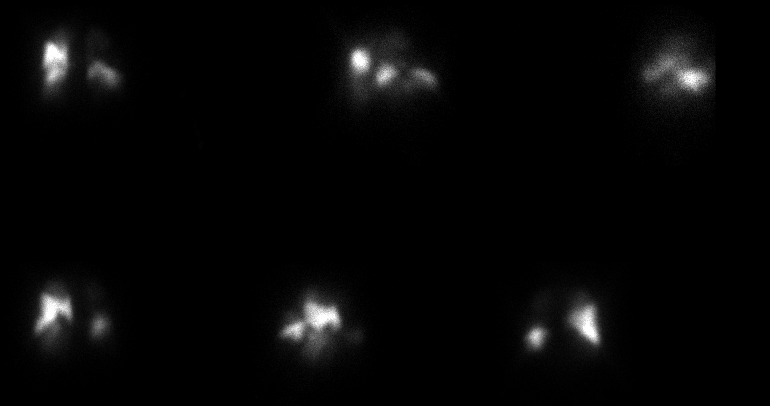
Pefusion images.
Top Row: Post, RPO, R lateral;
Bottom Row: LPO, L lateral, Anterior
View main image(pe) in a separate image viewer
View second image(vq).
Ventilation images. Sequential 1 minute frames.
The chest radiograph (not shown) was normal.
Full history/Diagnosis is available below
Diagnosis: Pulmonary embolism.
Full history:
Elderly woman who presented with acute gastrointestinal
bleeding approximately 3 weeks ago, now status post colectomy
who was apparently doing well postoperatively, with sudden onset of
tachypnea, decreased oxygen saturation, and decreased blood pressure.
Findings:
The ventilation study is normal.
The perfusion images are markedly abnormal, with nearly absent perfusion
to over 60% of the left lung and over 80% of the right lung. The
residual perfusion is noted in the superior segment of the right lower
lobe as well as a portion of the right middle lobe. On the left,
residual perfusion is to the anterior segment of the left upper lobe as
well as the posterior segment of the left upper lobe and lingula.
Discussion:
The markedly abnormal perfusion study, along with
a normal ventilation study and chest radiograph,
suggests massive pulmonary embolism. Even the areas
which are best perfused may not be completely free of
emboli, as partial occlusion may be present.
Given the patients hypotension, lytic agents should
at least be considered in the patient's management.
Followup:
An IVC filter was placed in the patient.
Major teaching point(s):
Within the "high" category, there is still a wide range
of scintigraphic appearance. This study has a very high
likelihood ratio for pulmonary embolism; both the level of
diagnostic certainty and the extent of disease should be
communicated to the referring physician.
ACR Codes and Keywords:
References and General Discussion of Ventilation Perfusion Scintigraphy (Anatomic field:Lung, Mediastinum, and Pleura, Category:Organ specific)
Search for similar cases.
Edit this case
Add comments about this case
Read comments about this case
Return to the Teaching File home page.
Case number: vq010
Copyright by Wash U MO

