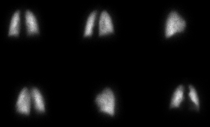
Pefusion images.
Top Row: Post, RPO, R lateral;
Bottom Row: LPO, L lateral, Anterior
View main image(pe) in a separate image viewer
View second image(vq). Ventilation images. Sequential 1 minute frames. (ignore the activity outside of the lungs on the single breath image)
The chest radiograph (not shown) was normal.
Full history/Diagnosis is available below
References and General Discussion of Ventilation Perfusion Scintigraphy (Anatomic field:Lung, Mediastinum, and Pleura, Category:Normal, Technique, Congenital Anomaly)
Return to the Teaching File home page.