Case Author(s): J. Wallis , 5/11/95 . Rating: #D1, #Q4
Diagnosis: Pulmonary embolism
Brief history:
Shortness of breath. Normal chest radiograph.
Images:

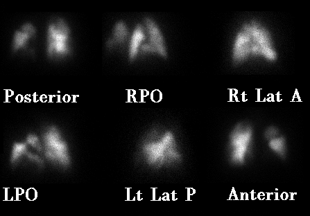
Perfusion images
View main image(vq) in a separate image viewer
View second image(vq).
Xe-133 ventilation images, one minute per frame
with washout images in the lower row.
The chest radiograph was normal.
Full history/Diagnosis is available below
Diagnosis: Pulmonary embolism
Full history:
The patient presented with recent onset of shortness of
breath and chest pain. There was no fever or cough
to suggest an infectious process.
Findings:
Multiple large defects are seen, including the
apical-posterior and anterior segments of the left
upper lobe, multiple left basilar defects, and a moderate
defect at the right apex.
Ventilation scintigraphy was normal, as was the chest
radiograph.
Discussion:
The findings easily meet the criteria for "high likelihood
ratio for pulmonary embolism" in both the modified Biello
and modified PIOPED schemes.
ACR Codes and Keywords:
References and General Discussion of Ventilation Perfusion Scintigraphy (Anatomic field:Lung, Mediastinum, and Pleura, Category:Organ specific)
Search for similar cases.
Edit this case
Add comments about this case
Read comments about this case
Return to the Teaching File home page.
Case number: vq008
Copyright by Wash U MO

