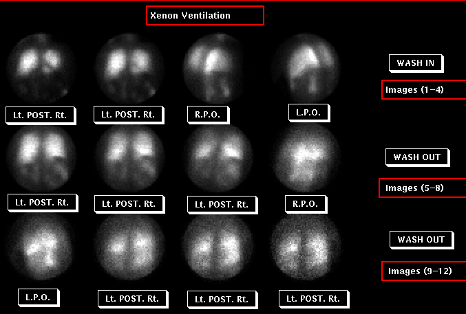
Xenon Ventilation Images
View main image(vq) in a separate image viewer
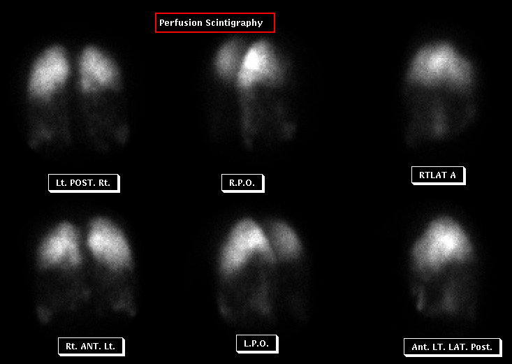
View second image(vq). Perfusion Images
Full history/Diagnosis is available below
Ventilation Xenon ventilation washin images demonstrate hypoventilation of the middle and lower lung zones bilaterally, with relative sparing of the upper lung zones. Washout images demonstrate corresponding areas of retension.
Perfusion: Perfusion images demonstrate a similar pattern, with marked hypoperfusion of the middle and lower lung zones bilaterally and relative sparing of both upper lung zones.
- Alpha-1 Antitrypsin is a glycoprotein synthesized in the liver and released in the serum.
- Injury occurs because the deficiency of alpha-1 antitrypsin prevents the neutralization of proteolytic enzymes released by alveolar macrophages and neutrophiils within the lung. These enzymes, especially elastase, destroy the basement membrane.
- Early Age of Onset (20 -30 years of age)
- M:F = 1:1
- Typical findings includ bullae at both lung bases with flattening of the diaphrams. Upper lung zones are generally unaffected.
(2) Quantitation revealed slightly greater ventilation and perfusion of the left lung compared to the right. Marked discrepancy would have affected the choice of which lung to replace, if unilateral lung transplantation were to be performed.
References and General Discussion of Ventilation Perfusion Scintigraphy (Anatomic field:Lung, Mediastinum, and Pleura, Category:Organ specific)
Return to the Teaching File home page.