Case Author(s): Hamid Latifi,MD and Jerold Wallis, MD , 8/22/94 . Rating: #D2, #Q3
Diagnosis: Right to left cardiac shunt
Brief history:
Pre lung transplantation workup
Images:
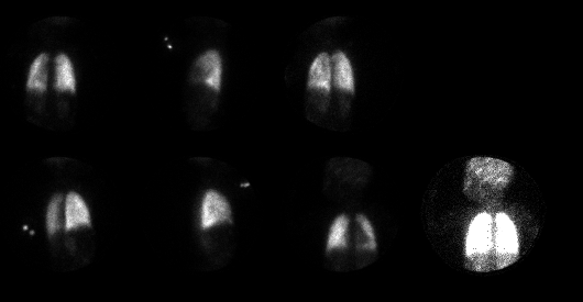
Above are posterior, left lateral, LPO,
RPO, right lateral and anterior images from
a perfusion scan. The anterior image is
shown a second time at higher intensity.
View main image(pe) in a separate image viewer
Full history/Diagnosis is available below
Diagnosis: Right to left cardiac shunt
Full history:
5 year old boy with slow growth, who was recently diagnosed
with atrial septal defect, patent ductus arteriosus, and
resultant pulmonary arterial hypertension. He is now being
evaluated for lung transplantation.
Radiopharmaceutical:
Tc-99m MAA, iv
Findings:
The perfusion images show accumulation of MAA in the kidneys
and brain, consistent with right to left shunt.
Differential Diagnosis List
Free pertechnetate can result in renal activity, but this pattern of activity in the brain is specific for a right-to-left shunt.
ACR Codes and Keywords:
References and General Discussion of Ventilation Perfusion Scintigraphy (Anatomic field:Heart and Great Vessels, Category:Normal, Technique, Congenital Anomaly)
Search for similar cases.
Edit this case
Add comments about this case
Return to the Teaching File home page.
Case number: vq003
Copyright by Wash U MO