Case Author(s): J. Philip Moyers, M.D. and Henry D. Royal, M.D. , 10/2/95 . Rating: #D1, #Q3
Diagnosis: Subacute testicular torsion
Brief history:
Testicular pain
Images:
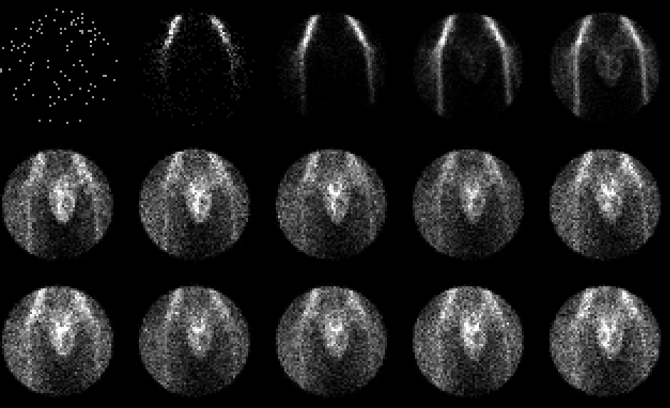
Anterior flow images of the scrotum
View main image(ts) in a separate image viewer
View second image(ts).
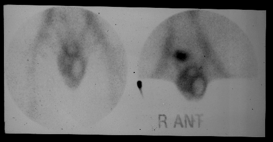
Immediate static images of the scrotum
Full history/Diagnosis is available below
Diagnosis: Subacute testicular torsion
Full history:
20 year old man who developed sudden left testicular pain three days prior to the scan. The pain has persisted over the three days, becoming more diffuse and dull in nature. He has noted left testicular swelling. The right testis is normal.
Radiopharmaceutical:
15.2 mCi Tc-99m pertechnetate i.v.
Findings:
The radionuclide angiogram
demonstrates increased blood flow in the region
surrouding the left testicle. The left testicle itself is
decreased in activity. The immediate static images
demonstrate no significant radiopharmaceutical
activity within the left testis although the tissue
surrounding the left testis demonstrates persistently
increased activity.
Discussion:
Scrotal imaging can be a useful
adjunct in the diagnosis of testicular torsion. At our
institution, if testicular torsion is strongly suspected,
the patient usually procedes to the operating room
without a diagnostic imaging study in order not to
delay diagnosis. Patients who are evaluated using
scrotal scintigraphy included those with a low pre-test
probability for testicular torsion in whom the
clinicians want to exclude testicular torsion and in
those with a late presentation or a confusing clinical
and physical findings. Scrotal imaging can be
valuable in these cases. The diagnosis of torsion
include acute and subacute torsion. Acute torsion is
defined as less than 24 hours. Testicular viability
decreases markedly after 24 hours and is best
preserved if the history of torsion is less than six
hours. In this case, a bulląs-eye appearance of an area
of decreased activity in the mid portion of the left
scrotum with a rim of increased activity suggests
subacute torsion. However, this appearance is not
pathognomonic and may be demonstrated with
abscess or hematoma. Epididymitis ususally has
increased blood flow and diffusely increased activity
on the immediate static images.
References: Mettler FA.
Essentials of Nuclear Medicine Imaging. 1991, 3rd
edition.
Followup:
The patient was referred to the
operating room. In the operating room, a 360 degree
torsion of the vascular supply to the left testicle and
an infarcted left testis was demonstrated. The left
testicle was removed. A right-sided orchiopexy was
performed.
Differential Diagnosis List
Abscess, hematoma, infected hydrocele
ACR Codes and Keywords:
References and General Discussion of Testicular Scintigraphy (Anatomic field:Genitourinary System, Category:Organ specific)
Search for similar cases.
Edit this case
Add comments about this case
Read comments about this case
Return to the Teaching File home page.
Case number: ts002
Copyright by Wash U MO