Case Author(s): J Lahorra MD and Keith Fischer MD , 6/21/95 . Rating: #D2, #Q4
Diagnosis: spermatic cord hematoma
Brief history:
pain in left testicle
Images:

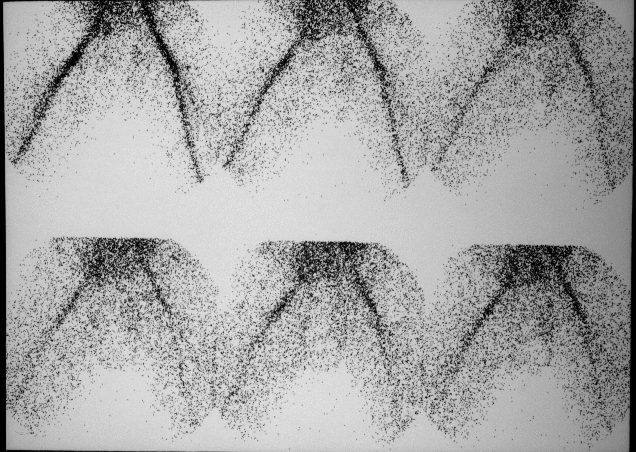
testicular scan- flow
View main image(ts) in a separate image viewer
View second image(ts).
testicular scan- immediate static images
View third image(us).
testicular ultrasound of the left testicle
Full history/Diagnosis is available below
Diagnosis: spermatic cord hematoma
Full history:
28 year old man with left testicular pain for several days.
No fevers.
Radiopharmaceutical:
Tc-99m Pertechnetate
Findings:
The testicular scan demonstrates no hyperperfusion on the flow study.
Immediate static images show a photopenic defect in the expected location of the left testicle with a rim of hyperemia. The ultrasound (actually obtained first), demonstrates a large heterogenous
mass filling the left hemiscrotum.
Discussion:
The testicular scan was obtained to confirm a possible tumor,
which would have been hyperemic on the flow portion of the study (even though US did not show the mass to be highly vascular). The diiferential diagnosis for a photopenic region with a hyperemic rim includes tumor with central necrosis (age of patient supports this), orchitis with abscess, infected hematoma, hydrocele, and late torsion.
Followup:
Surgery revealed a large, spermatic cord hematoma.
ACR Codes and Keywords:
References and General Discussion of Testicular Scintigraphy (Anatomic field:Genitourinary System, Category:Effect of Trauma)
Search for similar cases.
Edit this case
Add comments about this case
Return to the Teaching File home page.
Case number: ts001
Copyright by Wash U MO

