Case Author(s): Brock G. McDaniel, M.D. and Jerold Wallis, M.D. , 02/17/03 . Rating: #D2, #Q3
Diagnosis: Diffuse Metastatic Papillary Thyroid Carcinoma
Brief history:
60 year female with history of papillary thyroid carcinoma who is status post thyroidectomy and I-131 ablation 6 years ago.
Images:
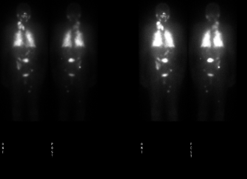
66 hour delay anterior and posterior images
View main image(tr) in a separate image viewer
View second image(xr).
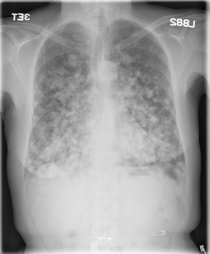
CXR
Full history/Diagnosis is available below
Diagnosis: Diffuse Metastatic Papillary Thyroid Carcinoma
Full history:
60 year old female with history of papillary thyroid carcinoma who is status post thyroidectomy and I-131 ablation of the residual thyroid tissue 6 years ago. The patient was then lost to follow-up until she presented to the emergency department 6 years later with complaints of acute shortness of breath and hemoptysis. Upon arrival to the emergency department she was found to have a heart rate of 128 bpm, a blood pressure of 102/54 mmHg, and an oxygen saturation of 91% on 5 liters of nasal cannula oxygen. A subsequent chest radiograph demonstrated diffuse pulmonary metastasis.
On physical examination a right neck mass was palpated. Fine needle aspiration confirmed recurrent follicular variant papillary thyroid carcinoma.
Radiopharmaceutical:
150 mCi therapeutic dose of I-131 sodium iodide, p.o.
Findings:
There is abnormal diffuse activity within the lungs bilaterally. There are at least three foci of increased uptake within the right neck and superior mediastinum, likely corresponding to recurrent thyroid carcinoma in the thyroid bed vs. metastatic lymphadenopathy. There are numerous additional metastatic lesions identified including a large focus in the mid abdomen, two foci in the pelvis - one within the posterior right pelvis and the other in the left hemipelvis, two foci within the left thigh, and a single focus in the distal right thigh.
The chest radiograph clearly demonstrates innumerable diffuse dense lung metastasis ranging in size from a few mm to 2.3cm.
Discussion:
Normally, I-131 activity can be seen in the choroid plexus, nasal mucosa/nasopharynx, salivary glands, thymus, stomach, bowel, urinary bladder, and breasts. Diffuse liver uptake may be seen on post-therapy scans and is diagnostic of thyroxine production. In post-ablation patients, liver activity suggests that I-131 has been incorporated into thyroid hormone by functioning metastases. A large amount of residual thyroid activity in the neck, however, may produce similar findings. It is interesting to note the lack of activity within the liver in this case. This is particularly surprising in this patient with diffuse pulmonary and soft tissue metastatic spread.
In women of childbearing age there is considerable uptake within lactating breasts, therefor, breast-feeding should be discontinued for at least 4-6 weeks prior to treatment in order to ensure mammary secretory activity has ceased. This will decrease the radiation dose to the breasts. Breast-feeding should be discontinued completely and not resumed following treatment. Breast uptake has been described in up to 6% of non-lactating females and may be asymmetric. Breast uptake may be confused with lung metastasis.
I-131 treatment prolongs survival in-patients with disseminated thyroid cancer, however, the rate of actual cure is low. I-131 ablation has been reported to be less effective in-patients younger than 40 years of age with metastases, however, these patients tend to live longer with metastatic disease.
In patients with lung metastases, a micronodular (miliary) pattern of metastases is associated with a good I-131 uptake and a better prognosis, while macronodular lesions (over 0.5 cm) frequently show poor uptake and have an associated worse prognosis. A rare complication in-patient with lung metastases is radiation-induced pneumonitis and pulmonary fibrosis. Given the extent of lung involvement in this patient, pneumonitis and pulmonary fibrosis must be considered. The risk can be decreased by choosing a treatment dose such that the whole body retention at 48 hours is less than 80 mCi.
ACR Codes and Keywords:
References and General Discussion of Thyroid Scintigraphy (Anatomic field:Face, Mastoids, and Neck, Category:Neoplasm, Neoplastic-like condition)
Search for similar cases.
Edit this case
Add comments about this case
Return to the Teaching File home page.
Case number: tr015
Copyright by Wash U MO