Case Author(s): Christian T. Schmitt, M.D. and Henry D. Royal, M.D. , 10/14/02 . Rating: #D3, #Q4
Diagnosis: Metastatic papillary thyroid carcinoma
Brief history:
74 year old man initially presented with right hip pain
Images:

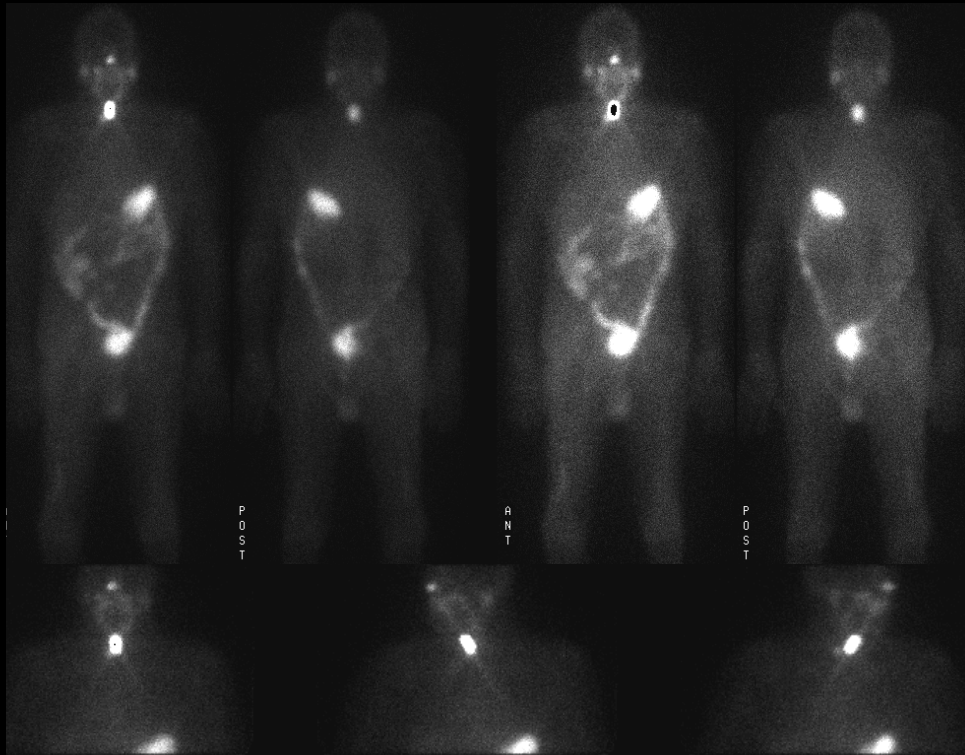
Anterior and posterior whole body and spot images are shown
View main image(tr) in a separate image viewer
View second image(pt).
Coronal FDG PET whole body images are shown
View third image(xr).
Chest radiograph is shown
View fourth image(ct).
CT of the chest is shown
Full history/Diagnosis is available below
Diagnosis: Metastatic papillary thyroid carcinoma
Full history:
74 year old man initially presented with right hip pain. Radiographs demonstrated a lytic lesion of the right acetabulum with a pathologic fracture. Core biopsy demonstrated metastatic papillary thyroid carcinoma. The patient underwent preoperative embolization of the right hip followed by total hip replacement. The patient also had thyroidectomy and received 100mCi of I-131. The initial images were from the subsequent whole body scans following I-131 treatment. The pathology from the thyroidectomy demonstrated metastatic papillary thyroid carcinom with anaplastic foci. The patient also had a PET scan for further evaluation.
Radiopharmaceutical:
First study: 100mCi I-131 sodium iodide, p.o.
Second study: 15mCi F-18 Fluorodeoxyglucose
Findings:
I-131 Whole Body Imaging: Increased uptake centrally in the neck consistent with residual functioning thyroid tissue, probably in the pyramidal lobe, no definite metastases.
Whole Body PET: Increased uptake within left jugular and supraclavicualr lymph nodes. Increased uptake within right paratracheal, subcarinal, right hilar lymph nodes and a right basilar lung nodule. Increased uptake in the right hip and pelvis. Moderate diffuse uptake in the bone marrow.
CXR: Right basilar pulmonary nodule and right hilar adenopathy.
Chest CT: Calcified right basilar pulmonary nodule and mediastinal lymphadenopathy.
Discussion:
This case demonstrates significant disseminated disease demonstrated on the PET study that was not identified on I-131 imaging. PET and I-131 imaging can be complementary in the evaluation of patients with thyroid cancer. PET is being employed currently in the follow up of patients with thyroid cancer with rising thyroglobulin levels after thyroidectomy, following a negative I-131 whole body exam.
View followup image(xr).
Pre- and post-surgical radiographs of the right hip
ACR Codes and Keywords:
- General ACR code: 23
- Face, Mastoids, and Neck:
2.3 "NEOPLASM, NEOPLASTIC-LIKE CONDITION"
References and General Discussion of Thyroid Scintigraphy (Anatomic field:Face, Mastoids, and Neck, Category:Neoplasm, Neoplastic-like condition)
Search for similar cases.
Edit this case
Add comments about this case
Return to the Teaching File home page.
Case number: tr014
Copyright by Wash U MO

