Case Author(s): J. Philip Moyers, M.D. and Farrokh Dehdashti, M.D. , 10/1/95 . Rating: #D1, #Q4
Diagnosis: Metastatic papillary thyroid carcinoma to the lungs and neck
Brief history:
Patient is status post
thyroidectomy for papillary carcinoma with follicular
variant.
Images:
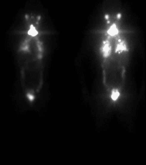
Anterior and posterior whole body I-131 images
View main image(tr) in a separate image viewer
Full history/Diagnosis is available below
Diagnosis: Metastatic papillary thyroid carcinoma to the lungs and neck
Full history:
18-year old woman with papillary
carcinoma of the thyroid status post total
thyroidectomy with removal of lymph nodes in the
neck and supraclavicular regions. MRI demonstrated
multiple pulmonary nodules.
Radiopharmaceutical:
200 mCi I-131 sodium iodide p.o.
Findings:
Anterior and posterior whole body I-
131 images demonstrate multiple areas of increased
uptake in the neck, as well as diffuse pulmonary
uptake.
Discussion:
Well differentiated papillary,
follicular, and mixed carcinomas are represented in
about 75% of all primary thyroid malignancies. The
overall 5-year survival rate of well differentiated
carcinoma is over 95% in properly treated patients.
Papillary carcinomas tend to metastasize via the
interstitium to local nodal groups while follicular
carcinomas tend to metastasize hematogenously. The
remaining thyroid malignancies include poorly
differentiated anaplastic thyroid carcinoma and
medullary carcinoma of the thyroid. Medullary
carcinoma of the thyroid may be seen in multiple
endocrine neoplasia type IIA and IIB. At our
institution, after a total or subtotal thyroidectomy, I-
131 whole body imaging is usually performed to
evaluate for metastatic disease. If metastatic disease
is demonstrated or if residual activity is demonstrated
within the neck, and it is unclear whether this
represents metastatic disease or functioning residual
thyroid tissue, the patient receives an ablative dose of
I-131. In this case, metastatic disease was suspected
on the basis of multiple pulmonary nodules
demonstrated on MRI. Therefore, an ablative dose
was given prior to a diagnostic dose and imaging was
performed after the ablative dose of 200 mCi of I-131.
To achieve maximum sensitivity in detecting
functioning lesions, follow-up whole-body I-131
imaging is usually performed 4-6 weeks after the
patient has been removed from thyroid
supplementation so that the patient has an elevated
TSH. Follow-up imaging may be done at 6 month
intervals (at shorter intervals in patients with
extensive or progressive disease and longer intervals
in patients with stable or slowly progressive disease)
until disease is eradicated and after which imaging
may be performed at 3-5 year intervals in patients
with high risk of recurrence. Since the dose is greater
than 30 mCi, the patient must be hospitalized
according to NRC regulations. In this case, multiple
small pulmonary nodules were demonstrated on MRI.
Other malignancies which can present as diffuse
multiple small pulmonary nodules include a GI
malignancy, usually pancreas, and GU malignancies,
usually renal cell carcinoma.
References:
1) Mettler FA. Essentials of Nuclear Medicine
Imaging. 1991 (3rd edition).
2) McDougall IR. Thyroid Disease in Clinical
Practice. New York, NY: Oxford University
Press, 1992 (1st edition).
Update 7/98. NRC regulations now allow larger doses to be
administered as an outpatient, provided one first documents the clearance
rate from the patient so that dosimetry to family members can
be calculated.
Followup:
The patient had MRI of the chest,
which showed multiple pulmonary nodules.
Major teaching point(s):
In order to diagnose
metastatic thyroid carcinoma, the normal
biodistribution of I-131 should be known. Normal
biodistribution of I-131 includes the salivary glands,
stomach, kidneys, and bowel. The lungs are not a
normal site of I-131 accumulation and if lung activity
is demonstrated, diffuse pulmonary metastatic
disease is the diagnosis.
ACR Codes and Keywords:
References and General Discussion of Thyroid Scintigraphy (Anatomic field:Face, Mastoids, and Neck, Category:Neoplasm, Neoplastic-like condition)
Search for similar cases.
Edit this case
Add comments about this case
Read comments about this case
Return to the Teaching File home page.
Case number: tr003
Copyright by Wash U MO